Brachycephalic Airway Obstruction Syndrome (BAOS)
in the Cavalier King Charles Spaniel
-
 What
It Is
What
It Is - Relation to Other Disorders
- Symptoms
- Care and Treatment
- Elongated Soft Palate
- Stenotic Nares
- Everted Laryngeal Saccules
- Laryngeal Collapse
- Pharyngeal Collapse
- Tracheal Collapse
- Sleep Disordered Breathing
- Breeders’ Responsibilities
- Research News
- Related Links
- Dedication
- Veterinary Resources
What It Is
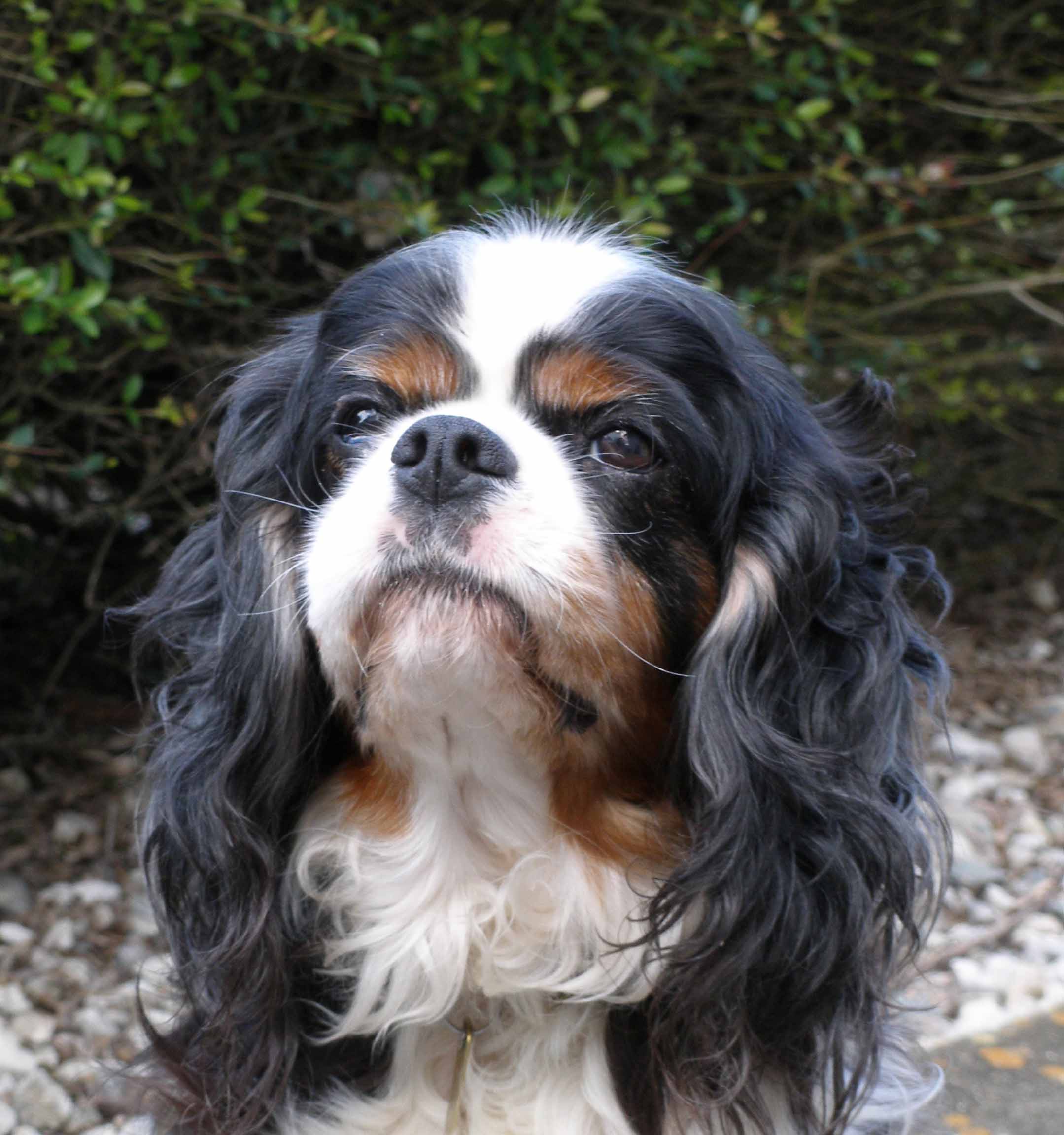
Brachycephalic airway obstruction syndrome (BAOS)* is characterized by primary and secondary upper respiratory tract abnormalities, which may result in significant upper airway obstruction. BAOS is an inherited condition in the cavalier King Charles spaniel. The breed is pre-disposed to it, due to the comparatively short length of the cavalier's head and a compressed upper jaw**. (Skull at right below is of a cavalier.)
* BAOS is also
referred to as brachycephalic airway disease (BAD) and brachycephalic airway
syndrome (BAS) and even brachycephalic obstructive airway syndrome (BOAS).
** Until 2011,
there had been some dispute among researchers as to whether the cavalier King
Charles spaniel is a brachycephalic breed or a mesaticephalic (or mesocephalic)
breed. However, in a
2011 German study, the researchers
concluded that the CKCS was brachycephalic but that it had a wider braincase in
relation to length than in other brachycephalic breeds.
 The term "brachycephalic" or "brachiocephalic" means short-headed and refers
to dogs with a shortened cranium (the bones that house the brain). Most
brachycepahic dogs have short muzzles and flat faces and noses which tip back
(airorhynchy) and a shorted lower jaw, but not necessarily.
The term "brachycephalic" or "brachiocephalic" means short-headed and refers
to dogs with a shortened cranium (the bones that house the brain). Most
brachycepahic dogs have short muzzles and flat faces and noses which tip back
(airorhynchy) and a shorted lower jaw, but not necessarily.
"Brachy" means short and "cephalic" means head. The throat and breathing passages in brachycephalic dogs often are undersized or flattened. The head's soft tissues are not proportionate to the shortened nature of the skull, and the excess tissues tend to increase resistance to the flow of air through the upper airway (nostrils, sinuses, pharynx and larynx).
This developmental defect is somewhat more apparent in a few other breeds: the English bulldog, pug, Boston terrier, and Pekingese, in particular. However, various degrees of BAOS predominate in the CKCS. The primary BAOS abnormalities in the cavalier include an elongated and fleshy soft palate, narrowed nostrils (stenotic nares), and everted laryngeal saccules, all of which are discussed in detail here. Other disorders may include laryngeal malformation and relatively small windpipe (tracheal hypoplasia or stenosis) and collapsing trachea*, and epiglottic retroversion, which are not specifically covered in this article. All of these disorders cause obstruction of the upper airway, compromise the dog's ability to take in air, and may result in laryngeal collapse. BAOS in the CKCS has been attributed by some researchers as a consequence of the selection for soft, puppy-like facial features, referred to as "morphological neoteny".
* Trachea collapse in the cavalier King Charles spaniel may also be due to an enlarged heart caused by advanced mitral valve disease. Also, a 2004 study by researchers at the Royal Veterinary College found that 53% of the brachycephalic dogs in their 92 dog sample had heart disease, compared to 24% of the non-brachycephalic dogs. See Mitral Valve Disease.
In a 2010 report of BAOS surgery on 155 Australian dogs, the cavalier was the most common breed (29 dogs, 18.7%). The researchers found: "All CKCS had an elongated soft palate and accounted for 41% of the laryngeal collapse cases."
RETURN TO TOP
Relation to Other Disorders
In a 2010 report, UK researchers found an association between primary secretory otitis media (PSOM) and brachycephalic conformation in cavaliers. They stated: "In CKCS, greater thickness of the soft palate and reduced nasopharyngeal aperture are significantly associated with OME [otitis media with effusion, meaning PSOM]." However, they did not explain why PSOM is so nearly limited to the cavalier, while so many other breeds are brachycephalic. They also concluded that bilateral PSOM was associated with CKCS with more extreme nasopharyngeal conformation, than unaffected CKCS.
Robert N. White, a board certified veterinary soft tissue surgeon practicing at Willows Veterinary Centre and Referral Service in Solihull, West Midlands, observed in an October 2010 presentation before a meeting of the UK's Association of Veterinary Soft Tissue Surgeons, that the cavalier King Charles spaniel does not appear to be a classically brachycephalic breed, despite the extent of BAOS in the cavalier, and that the extent of both primary secretory otitis media (PSOM) and syringomyelia (SM) in the breed suggests that the CKCS may suffer from a combination syndrome of the three disorders, all associated with Chiari-like malformation.
In a January 2017 article, UK researchers found evidence that pain resulting from Chiari-like malformation (CM) is associated with increased brachycephaly, and syringomyelia (SM) can result from different combinations of abnormalities of the forebrain, caudal fossa, and craniocervical junction which compromise the neural parenchyma and impede cerebrospinal fluid flow.
In a December 2022 article, ophthalmologists attribute the high prevalence of both dry eye syndrome and corneal ulcers in cavaliers to BAOS.
RETURN TO TOP
Overall Symptoms
The symptoms may vary and range in severity, depending upon which abnormality is causing them, but they typically include:
• labored and constant open mouthed breathing
• noisy breathing
• snuffling
• snorting
• excessive or loud snoring
• gagging
• retching
• sleep apnea
• exercise and/or heat intolerance
• general lack of energy
• pale or bluish tongue and gums due to a lack of oxygen
Audible breathing noises when inhaling, such as a high-pitched (stridor) or low pitched noise (stertor) likely is symptomatic of upper respiratory tract disease. Stridor, the high-pitched noise, is associated with upper airway obstruction and disease of the oropharynx or larynx. Stertor, the lower-pitched noise, is similar to snoring and would be associated with disease of the nasal cavity or nasopharynx.
In an April 2018 abstract, a team of UK veterinary surgeons reported their study of five cavaliers which had been suffering from obstructive sleep apnea (apnoea). Apnea means the suspension of breathing due to blocked airways. Previously, it had been reported only in English bulldogs, French bulldogs, and pugs, all typically severely brachycephalic breeds. All five CKSCs suffered from heavy snoring and noisy breathing and choking, as well as sleep apnea. In addition, all five had eosinophilic stomatitis and primary secretory otitis media (PSOM); four had mitral valve disease (MVD); and three had syringomyelia (SM). Also, all five had "aberrant nasal turbinates, nasal septal deviation and soft palate thickening and 3/5 had nasopharyngeal thickening and tracheal collapse."
Also, studies have concluded that brachycephalic dogs may be predisposed to the conditions of Primary Secretory Otitis Media (PSOM), and eye problems, such as entropion, dry eye, and other disorders which may be caused by eyes not properly seated in the skull.
RETURN TO TOP
Overall Care and Treatment
The brachycephalic cavalier is an inefficient panter. Increased panting can cause swelling and narrowing of the airway, resulting in collapsing or fainting. The excessive panting and episodes of other symptoms may place increased strain on the dog's heart, which cavaliers with mitral valve disease cannot afford to risk.
Care should be taken to avoid overheating and excessive excitement and excessive exercise, which may cause increased panting. Excessive barking or panting may cause the throat to swell, which could result in a totally blocked airway. Most importantly, the owner should not let the dog get too hot, particularly in the summer months, and not allow the dog to become overweight, as obesity will exacerbate the respiratory difficulties. Death from such related causes as heat stroke may be the consequence of not diagnosing and treating these symptoms early enough.
 Care also should be taken when anesthesia may have to be
administered. In a March 2012 report
by a team of Tufts University veterinary anesthesiologists, cavaliers are among
brachycephalic breeds which require special attention when being sedated and
anesthetized. Their advice includes:
Care also should be taken when anesthesia may have to be
administered. In a March 2012 report
by a team of Tufts University veterinary anesthesiologists, cavaliers are among
brachycephalic breeds which require special attention when being sedated and
anesthetized. Their advice includes:
"Avoid excessive sedation. Avoid α2-agonists. Administer acepromazine at half dose. Preoxygenate. Use short-acting induction agent. Use appropriately sized endotracheal tubes. Extubate after patient is sitting up, vigorously chewing, bright, alert."
See also our webpage on anesthesia and sedatives.
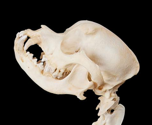
In mild episodes, calming and cooling the dog may be sufficient. Inflammation and swelling of the airway tissues (oedema) may be treated with oxygen therapy (see photo) and corticosteroids for short term relief. Surgery is required when the abnormalities chronically interfere with breathing. Following surgery, the dog will need to be monitored closely for at least the first 24 hours, because inflammation or bleeding can obstruct the airway, making breathing difficult or impossible.
In all cases, it is strongly recommended that only board certified veterinary surgeons (who also are very experienced at airway surgery) be permitted to perform any type of airway surgery on cavaliers.
 Unfortunately, the only solution to most cases of BAOS is some form
of surgery. In a
June 2016 article, the Spanish specialist definitively states:
Unfortunately, the only solution to most cases of BAOS is some form
of surgery. In a
June 2016 article, the Spanish specialist definitively states:
"The definitive treatment for the BS [brachycephalic syndrome] is always surgical. Early intervention can slow down the progression of the signs and complications. There are several surgical techniques and, during the last years, the use of CO2 laser ensures less surgical time, bleeding, swelling and intra and postoperative pain, as well as a more precise tissue ressection. A possible explanation for the poor therapeutic success after conventional surgeries, could be the lack of consideration in the diagnosis, management and treatment of the rest of intranasal structures. To understand the BS, all the efforts should relapse into the selection of these breeds, so that in the future, the objective is to encourage more moderate craniofacial morphologies in order to reduce the prevalence and severity of BS."
RETURN TO TOP
Elongated & Fleshy Soft Palate -- Reverse Sneeze
-- what it is
 The
palate is the roof of the mouth. It is divided into two parts, the anterior
(front) bony
hard palate, and the posterior (rear) fleshy soft palate. The soft palate separates the
nasal passage from the oral cavity. An elongated soft palate is too long for the
length of the mouth, so that its tip protrudes into the front of the airway and
may get sucked into the laryngeal opening where it may obstruct the normal
passage of air into the trachea. A fleshy soft palate is an
abnormally thick one which reduces the dimension of the nasal air passage way.
(See the soft palate at the top of the sketch, right.)
The
palate is the roof of the mouth. It is divided into two parts, the anterior
(front) bony
hard palate, and the posterior (rear) fleshy soft palate. The soft palate separates the
nasal passage from the oral cavity. An elongated soft palate is too long for the
length of the mouth, so that its tip protrudes into the front of the airway and
may get sucked into the laryngeal opening where it may obstruct the normal
passage of air into the trachea. A fleshy soft palate is an
abnormally thick one which reduces the dimension of the nasal air passage way.
(See the soft palate at the top of the sketch, right.)
RETURN TO TOP
-- symptoms
The most common and recurrent symptom of an elongated or
fleshy soft palate is noisy
breathing. Occasionally, the dog will make snorting sounds, which is due to the
tip of the palate flapping into the trachea
 during respiration. This is called
the "Cavalier snort" or a "reverse sneeze".* The dogs also are more likely to
snore, gag, or retch, and in severe instances, they may collapse if the airflow
is obstructed completely. See an example of a cavalier reverse sneezing at
this
YouTube video. See also, our blog entry,
All that cavalier owners need to know about the “Reverse Sneeze” or “Cavalier
Snort”.
during respiration. This is called
the "Cavalier snort" or a "reverse sneeze".* The dogs also are more likely to
snore, gag, or retch, and in severe instances, they may collapse if the airflow
is obstructed completely. See an example of a cavalier reverse sneezing at
this
YouTube video. See also, our blog entry,
All that cavalier owners need to know about the “Reverse Sneeze” or “Cavalier
Snort”.
* It may be confused with pharyngeal gag reflex or inspiratory proxysmal respiration.
Reverse sneezes may occur due to any of a variety of causes, such as foreign bodies, airborne irritation, or growths in the nasal passage. These possible causes need to be eliminated during the diagnosis procedures.
Cavaliers with abnormally thick soft palates also are more likely to develop primary secretory otitis media (PSOM), due to the size of the soft palate impairing auditory tube drainage.
RETURN TO TOP
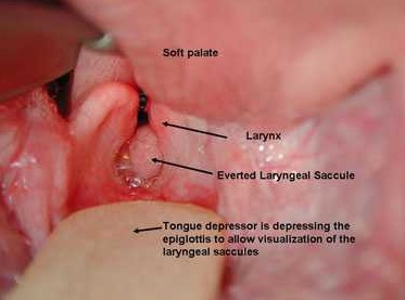
-- diagnosis
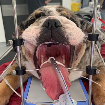
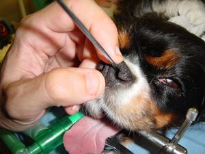
In severe cases, the palate usually is examined with the dog under light general anesthesia, using a laryngoscope. An elongated palate will obstruct the view of the larynx when the tongue is depressed. The veterinarian may take an x-ray to determine the length of the palate and airway.
RETURN TO TOP
-- treatment
 If the palate is only moderately elongated and does not totally block the
trachea, most cavaliers are able to pull out of these blockages by themselves. Snorting may be relieved by forcing the cavalier to breathe through its
mouth instead of its nose. This may be done by holding the dog's head down and
mouth open with one hand while blocking air from entering the nose with the
other hand.
If the palate is only moderately elongated and does not totally block the
trachea, most cavaliers are able to pull out of these blockages by themselves. Snorting may be relieved by forcing the cavalier to breathe through its
mouth instead of its nose. This may be done by holding the dog's head down and
mouth open with one hand while blocking air from entering the nose with the
other hand.
Treatment for recurring blockage of airflow is surgical removal of excess tissue from the palate by a veterinary surgeon. One of those procedures is "folded flap palatoplasty" (FFP), which both shortens and thins the soft palate. Others are carbon dioxide laser for resectioning, and harmonic scalpels. See this November 2011 article and this October 2017 article for additional details.
Post surgery prognosis is good for young dogs. They generally may be expected to breathe much easier, with significantly reduced respiratory distress, and display more energy and stamina. Older dogs may have a less favorable prognosis. However, in this November 2011 article, there is this report of a very successful 5 minute surgery on a 7 year old CKCS:
"A 7-year-old male neuter Cavalier King Charles Spaniel was presented with moderate respiratory compromise secondary to BAOS. The patient had a history of slowly worsening inspiratory obstruction and was becoming increasingly exercise intolerant. The nares were not stenotic and the laryngeal saccules appeared normal. Under general anaesthesia it was judged that the soft palate was 5 mm too long and surgery was performed to resect the redundant tissue. The staphylectomy took 5 min without complications. The modified surgical procedure was used in this case and the patient recovered uneventfully, with no postoperative bleeding or respiratory compromise. Respiratory function was much improved at recovery from anaesthesia and at 24 h. The patient was virtually asymptomatic 6 months postoperatively."
In all cases, it is strongly recommended that only board certified veterinary surgeons (who also are very experienced at airway surgery) be permitted to perform any type of airway surgery on cavaliers.
In a 2010 report of BAOS surgery on 155 Australian dogs, the cavalier was the most common breed (29 dogs, 18.7%). All of those cavaliers had an elongated soft palate.
RETURN TO TOP
Stenotic Nares
-- what they are
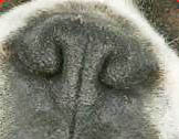 Stenotic nares are abnormally narrow or obstructed nostrils
(right), especially when
inhaling. Dogs with this disorder tend to breathe primarily through their
mouths, because breathing through the nose is unproductive. When they do breathe
through their noses, they make wheezing sounds. Stenotic nares cause the dog to
inhale deeper to draw air through the nose and into the lower airway, which may
contribute to the development of secondary abnormalities, such as
everted laryngeal saccules and
laryngeal collapse.
Stenotic nares are abnormally narrow or obstructed nostrils
(right), especially when
inhaling. Dogs with this disorder tend to breathe primarily through their
mouths, because breathing through the nose is unproductive. When they do breathe
through their noses, they make wheezing sounds. Stenotic nares cause the dog to
inhale deeper to draw air through the nose and into the lower airway, which may
contribute to the development of secondary abnormalities, such as
everted laryngeal saccules and
laryngeal collapse.
RETURN TO TOP
-- symptoms
The dog's nose appears narrow, with the nostril wings (alar folds) collapsing inward during inhalation and possibly blocking the nares. As noted above, the dog tends to breathe through its mouth, and makes wheezing sounds when breathing with its mouth closed. Symptoms typically include labored and constant open mouthed breathing, noisy breathing, snuffling, snorting, excessive snoring, and in severe cases, gagging, retching, exercise and/or heat intolerance, pale or bluish tongue and gums due to a lack of oxygen.
RETURN TO TOP
-- diagnosis
Stenotic nares are easily diagnosed by visual examination. In severe cases, the flow of air through the nostrils may be so poor that no air movement can be detected.
RETURN TO TOP
-- treatment
 Surgery under general anesthesia is the preferred means of treating stenotic
nares. Surgical techniques for widening the nares include: alapexy, punch
resection, partial amputation of the nasal cartilage, dorsal offset rhinoplasty,
and multiple variations of wedge excision of the ala nasi. The aim is to increase the size of the nostrils by removing tissue from
the wings and possibly some related cartilage.
Surgery under general anesthesia is the preferred means of treating stenotic
nares. Surgical techniques for widening the nares include: alapexy, punch
resection, partial amputation of the nasal cartilage, dorsal offset rhinoplasty,
and multiple variations of wedge excision of the ala nasi. The aim is to increase the size of the nostrils by removing tissue from
the wings and possibly some related cartilage.
In an August 2020 article, USA surgeons report surgically repairing the stenotic nares of 34 brachycephalic dogs, including one cavalier, using the dorsal offset rhinoplasty (DOR) technique. DOR involves removing a wedge of nasal planum and cartilage from each nare, opening the nares.
Post surgery recovery is similar to that described above under Overall Care and Treatment and treatment for elongated soft palate. Following surgery, the dog will be required to wear an Elizabethan collar to keep the surgical site clean and to protect it from rubbing.
In all cases, it is strongly recommended that only board certified veterinary surgeons (who also are very experienced at airway surgery) be permitted to perform any type of airway surgery on cavaliers.
RETURN TO TOP
Everted Laryngeal Saccules
-- what they are
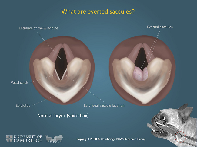 Everted laryngeal saccules are a secondary abnormality to either an elongated
soft palate or stenotic nares. The larynx contains the vocal chords, produces
sound, and protects the trachea. It is located at the point where the upper
tract divides into the trachea and the esophagus. During swallowing, the larynx
closes to prevent swallowed material from entering the lungs. The laryngeal
saccules are part of the mucosal lining of the laryngeal ventricles and appear
as two membranous sacs that are located in recessions in front of the vocal
folds.
Everted laryngeal saccules are a secondary abnormality to either an elongated
soft palate or stenotic nares. The larynx contains the vocal chords, produces
sound, and protects the trachea. It is located at the point where the upper
tract divides into the trachea and the esophagus. During swallowing, the larynx
closes to prevent swallowed material from entering the lungs. The laryngeal
saccules are part of the mucosal lining of the laryngeal ventricles and appear
as two membranous sacs that are located in recessions in front of the vocal
folds.
Brachycephalic cavaliers must create more pressure when they inhale in order to fill their lungs with air. This decreases the pressure in the upper airway, causes the lining of the larynx (laryngeal ventricles) to swell, and forces the laryngeal saccules to vibrate and evert into the airway at the opening to the trachea, blocking the flow of air. Everted laryngeal saccules usually are the first stage of laryngeal collapse.
RETURN TO TOP
-- symptoms
The symptoms are those common to a lack of air intake, such as gagging, retching, fainting, pale or bluish tongue and gums due to a lack of oxygen.
RETURN TO TOP
-- diagnosis
Everted laryngeal saccules are diagnosed under anesthesia. They appear as bilateral, red, fleshy, globular sacs.
RETURN TO TOP
-- treatment
Tissue from the saccules are surgically removed under general anesthesia. This procedure is called a “laryngeal sacculectomy”. A tube will be inserted through the neck into the trachea (“temporary tracheostomy”) to allow an airway during surgery and will remain until the swelling in the throat subsides enough that the dog can breathe normally. Post surgery and recovery are similar to that described above under under Overall Care and Treatment and treatment for elongated soft palate.
In all cases, it is strongly recommended that only board certified veterinary surgeons (who also are very experienced at airway surgery) be permitted to perform any type of airway surgery on cavaliers.
RETURN TO TOP
Laryngeal Collapse
-- what it is
Laryngeal collapse is an advanced form of brachycephalic airway obstruction syndrome.* The primary conditions of stenotic nares and elongated soft palate, together with everted laryngeal saccules, lead to abnormal stresses on the larynx and a progressive distortion and ultimate collapse of the cartilage supporting the larynx. There are three stages (grades) of laryngeal collapse, Stage 1 (Grade 1) being the everted laryngeal saccules described above. Stage 2 (Grade 2) occurs when the arytenoid cartilage loses its rigidity and gradually collapses inwardly. The third and final stage (Grade 3) is when the cartilage fails completely, and the larynx collapses.
* A similar condition is tracheal collapse, which appears less common in the CKCS. Also, laryngeal paralysis.
In a 2010 report of BAOS surgery on 155 Australian dogs, the cavalier was the most common breed (29 dogs, 18.7%). The researchers found: "All CKCS had an elongated soft palate and accounted for 41% of the laryngeal collapse cases."
RETURN TO TOP
-- symptoms
When the larynx collapses, the cavalier will not be able to breathe at all. The situation will be an extreme, life-threatening emergency.
RETURN TO TOP
-- diagnosis
The diagnosis is made by visual examination using a laryngoscope while the dog is under light anesthesia. A laryngoscope is a thin tube with a light, lens, and a video camera. Radiiography (X-rays) of the laryngeal ventricles is being researched as another diagnositic device. It involves measuring the ratios of the ventricular length and surface to the length of the third cervical vertebra (MVL/LC3 and VS/LC3).
RETURN TO TOP
-- treatment
Depending upon the severity of the collapse, an early option may be a partial laryngectomy to enlarge the laryngeal opening. The procedure will include anesthesia and a temporary tracheostomy. Statistical studies have shown that less than 50% of dogs treated this way survive, due to the permanent cartilage deformation and softening which results in continued collapse.
Permanent tracheostomy is the only other option. A permanent tracheostomy creates a permanent opening (a stoma) through the neck into the trachea (windpipe). A tracheostomy tube or trach tube is placed through the stoma to provide an airway and to remove secretions from the lungs. Prognosis is poor, particularly for older dogs.
In a June 2018 article about permanent tracheostomies performed on 15 brachycephalic dogs with severe laryngeal collapse, a team of Italian veterinary surgeons (Matteo Gobbetti, Stefano Romussi, Paolo Buracco, Valerio Bronzo, Samuele Gatti, Matteo Cantatore) reported on three surgeries required to resolve grade 3 laryngeal collapse in a cavalier King Charles spaniel. The CKCS was an 11 year old female. The first procedure shorted the dog's elongated soft palate. A month later, the surgeons resected the cavalier's hypertrophic aryepiglottic folds surrounding the larynx. A month after the second procedure, the surgeons performed a permanent tracheostomy, which creates a permanent opening (a stoma) through the neck into the trachea (windpipe). A tracheostomy tube or trach tube is placed through the stoma to provide an airway and to remove secretions from the lungs. The CKCS survived the three surgeries successfully and lived another 143 days, dying by euthanasia due to a separate medical disorder.
A 2014 report examined the insertion of silicone tracheal stoma stents for temporary tracheostomy in eighteen dogs with upper airway obstruction, including three cavaliers. The conclusion was that use of the stent beyond five days was not recommended because of granulation tissue formation, and that the long-term consequences of partial tracheal ring resection are unknown.
In all cases, it is strongly recommended that only board certified veterinary surgeons (who also are very experienced at airway surgery) be permitted to perform any type of airway surgery on cavaliers.
RETURN TO TOP
Pharyngeal Collapse
-- what it is
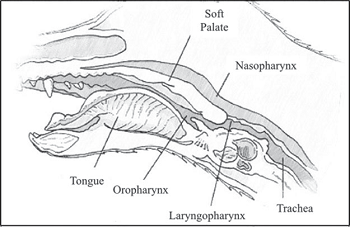 The
pharynx is the dog's throat, a muscular membrane tube that extends from
the soft palate at the back of the tongue and nasal cavity and larynx
down to the where its trachea and esophagus come together. It has three
regions, the nasopharynx at its top, the oropharynx at its center behind
the mouth, and the laryngopharynx at its bottom. The nasopharynx is
connected to the nasal cavities and is the air passage during breathing
and also connects to the openings of the eustachian tubes through which
air enters the middle ear. The oropharynx and laryngopharynx are
passageways for both air and food. (See diagram from
Rubin et al., at right.)
The
pharynx is the dog's throat, a muscular membrane tube that extends from
the soft palate at the back of the tongue and nasal cavity and larynx
down to the where its trachea and esophagus come together. It has three
regions, the nasopharynx at its top, the oropharynx at its center behind
the mouth, and the laryngopharynx at its bottom. The nasopharynx is
connected to the nasal cavities and is the air passage during breathing
and also connects to the openings of the eustachian tubes through which
air enters the middle ear. The oropharynx and laryngopharynx are
passageways for both air and food. (See diagram from
Rubin et al., at right.)
When pharyngeal collapse occurs, the tube is weakened and narrows, particularly at the nasopharynx. It may occur upon inhaling or upon exhaling, depending upon the underlying cause. Pharyngeal collapse is not believed to be a primary disease and instead a complication of other airway disorders. Negative airway pressure upon inhalation and increased pressure upon exhalation, due to the underlying primary disorder, can cause the pharynx to collapse, either partially or completely. In general, brachycephalic dogs suffering from an elongated soft palate or everted laryngeal saccules or stenotic nares, can affect pharyngeal diameter.
RETURN TO TOP
-- symptoms
The most common sign is coughing, followed by snoring, gagging, regurgitation of food, and sneezing. Sleep disordered breathing, in particluar apnea, may also be attributed to pharyngeal collapse upon exhaling.
RETURN TO TOP
-- diagnosis
Fluoroscopy (or videofluoroscopy), which shows a continuous x-ray image, so that the pharynx can be viewed in motion upon inhaling and exhaling, can confirm pharyngeal collapse. The dog is awake during this procedure, although it may be sedated. Computed tomography (CT) scans can assess structural abnormalities in the pharynx.
RETURN TO TOP
-- treatment
Since pharyngeal collapse usually is secondary to an underlying disorder, treatment usually is of that primary cause.
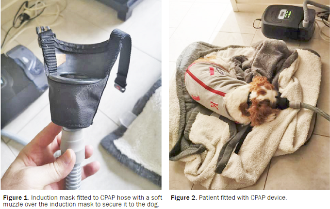
In an April 2021 article, a 4-year-old female cavalier King Charles spaniel diagnosed with partial pharyngeal collapse experienced sleep apnea episodes every 10 to 15 minutes during sleep. The dog was fitted with a "continuous positive airway pressure" (CPAP) device, which included a mask connected to an air hose and pump, to create a mild positive pressure in the dog's airway. (See Figures 1 & 2.) A muzzle was fitted over the mask to secure it to the dog's head. The cavalier gradually got accustomed to the device, with the owner using positive reinforcement with treats and removing the apparatus before the dog displayed signs of distress. Eventually, after continuing to suffer SDB episodes without the device, the dog sought out the mask and muzzle. With the CPAP device in place, the cavalier was able to sleep between 4 to 5 hours each night.
RETURN TO TOP
Tracheal Collapse
 -- what it is
The trachea is a flexible tube that connects the larynx with the mainstem bronchi. The tube is composed of 35 to 45 C-shaped rings of cartilage, held by the trachealis muscle and membranes. The trachea is commonly referred to as the windpipe. The interior airspace of the trachea is called the tracheal lumen. The trachea transports air through its lumen to and from the lungs during respiration and also transports debris to the larynx.
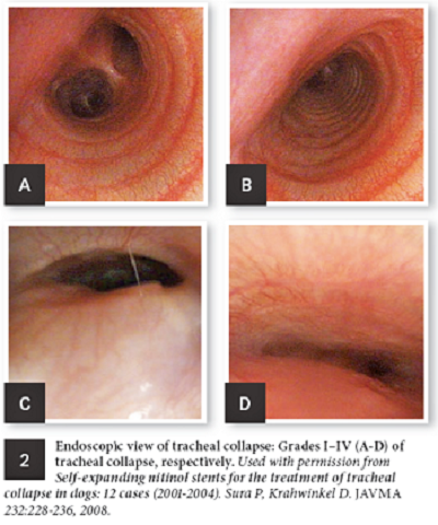
Tracheal collapse is a progressive flattening of the tracheal lumen. It begins with the trachealis muscle weakening and progresses to the C-shaped rings becoming more egg-shaped, resulting in the lumen to narrow and flatten. (See set of endoscopic views at right.)
The severity of tracheal collapse is graded based upon the percentage of blockage of the lumen: from Grade I (<25%), to Grade II (<50%) to Grade III (<75%) to Grade IV (up to 100% or complete blockage).
RETURN TO TOP
-- symptoms
The most common sign is coughing, particularly a goose-like honking, noisy breathing, then difficulty breathing, bluish discoloration of the tongue and/or gums, and high body temperature. The trachea changes postion, its cartilege may make a clicking sound when the dog breathes.
Note that in cases of cavaliers with mitral valve disease (MVD) with heart enlargement, the enlarged left atrium (LA) may press against the trachea and cause the dog to cough as if the trachea has collapsed.
RETURN TO TOP
-- diagnosis
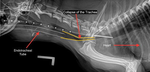 X-rays
are the quickest and least expensive method of detecting a
collapsing trachea. (See x-ray at right from the
Univ. of Missouri.)
X-rays
are the quickest and least expensive method of detecting a
collapsing trachea. (See x-ray at right from the
Univ. of Missouri.)
Fluoroscopy (or videofluoroscopy), which shows a continuous x-ray image, so that the trachea can be viewed in motion upon inhaling and exhaling, can confirm tracheal collapse. The dog is awake during this procedure, although it may be sedated. Computed tomography (CT) scans can assess structural abnormalities in the trachea. Other methods include ultrasound and tracheobronchoscopy.
RETURN TO TOP
-- treatment
Most dogs having tracheal collapse are medicated, primarily with a cough suppressant/antitussive (diphenoxylate hydrochloride and atropine sulfate [Logen, Lomotil, Lonox, Lofenoxal], hydrocodone, butorphanol, codeine phosphate) to reduce the frequency of coughing. Corticosteroids, such as stanozolol, my be added to reduce inflammation resulting from the collapse. Secondary airway infections will require antibiotics (doxycycline, cephalexin, or amoxicillin–clavulanate). Bronchodilators (theophylline, albuterol* [salbutamol] and , terbutaline, aminophylline) and/or antihistamines may also be prescribed.
* Albuterol (salbutamol) reportedly is toxic to some dogs, especially the aerosolized version. Toxicity symptoms have included tachycardia or other arrhythmia, weakness, lethargy, tachypnea, aggression, agitation, hyperactivity, vomiting, tremors and seizures.
More severe cases will require tracheal stent implantation inside the lumen. Stents are woven and self-expanding, made of the nickel-titanium alloy nitinol. The stent is inserted by means of a catheter.
Overweight dogs may be expected to benefit from weight loss. Neck collars and neck leads should be avoided, and instead a harness should be used when walking the dog, to avoid pressure acraoss the neck. Difficulty breathing and/or a bluish tongue will require oxygen supplementation, injected corticosteroids, and sedatives.
RETURN TO TOP
Sleep Disordered Breathing
Sleep disordered breathing (SDB) refers to any breathing difficulties which occur while the dog is asleep. They range from snoring due to partial obstructions to apena, which is the complete cessation of breathing. In cavalier King Charles spaniels, it most often is attributable to BAOS.
In a February 2024 article, a team of Finnish researchers evaluated the risk factors for sleep-disordered breathing (SDB) in serveral dog breeds. Of 28 brachycephalic dogs, 8 (28.5%) were cavalier King Charles spaniels. Other SDB breeds comprised 35 dogs. They reported that "We had an overrepresentation of Cavalier King Charles Spaniels, French Bulldogs and Labrador Retrievers." They found that brachycephaly was a significant risk factor for SDB, particularly apnea, and BOAS-positive class (moderate or severe BOAS signs) was a significant risk factor, along with excess weight. Aging and Chiari-like malformation were not risk factors among the dogs in this study.
• Apnea
Apnea means the complete suspension of breathing due to blocked airways. In dogs, it also is called obstructive sleep apnea because it usually is attributed to obstruction of the brachycephalic dog's upper airway, a sign of brachycephalic airway obstruction syndrome (BAOS). This form of apnea is most common among English bulldogs, French bulldogs, and pugs, all typically severely brachycephalic breeds, but has been observed in cavalier King Charles spaniels as well.
Symptoms of apena include interrupted sleep, choking, and other signs
that the dog has ceased to breathe. The dog may wake up in distress,
gasping. It may sleep with its chin elevated or mouth open to compensate
for the
disorder.
in most cases, the cavalier has been diagnosed with nasal septum
deviation (NSD). The septum is comprised of bone and cartilage,
which divides the right and left nostrils (nares) and airways of the
nasal cavity. Deviation of the septum means that the septum is misplaced
and one or both of the nostrils is abnormally small. See this
March 2022 article.
In severe cases, the treatment option is limited to surgery of the portion of the airway causing the obstruction, typically at the nasal entrance, nasal cavity, pharynx, and/or larnynx. (See photo.)
In an April 2018 abstract and a May 2019 article, a team of UK veterinary surgeons reported their study of five cavaliers which had been suffering from obstructive sleep apnea (apnoea). All five CKSCs suffered from heavy snoring and noisy breathing and choking, as well as sleep apnea. In addition, all five had eosinophilic stomatitis and primary secretory otitis media (PSOM); four had mitral valve disease (MVD); and three had syringomyelia (SM). Also, all five had "aberrant nasal turbinates, nasal septal deviation and soft palate thickening and 3/5 had nasopharyngeal thickening and tracheal collapse."
In an April 2021 article, a 4-year-old female cavalier King Charles spaniel diagnosed with partial pharyngeal collapse experienced sleep apnea episodes every 10 to 15 minutes during sleep. The dog was fitted with a "continuous positive airway pressure" (CPAP) device, which included a mask connected to an air hose and pump, to create a mild positive pressure in the dog's airway. (See Figures 1 & 2.) A muzzle was fitted over the mask to secure it to the dog's head. The cavalier gradually got accustomed to the device, with the owner using positive reinforcement with treats and removing the apparatus before the dog displayed signs of distress. Eventually, after continuing to suffer SDB episodes without the device, the dog sought out the mask and muzzle. With the CPAP device in place, the cavalier was able to sleep between 4 to 5 hours each night.
See, also, our discussion of stenotic nares.
RETURN TO TOP
• Snoring
 Snoring is not normal for dogs, particularly loud snoring. It is a
symptom of sleep disordered breathing (SBD) caused by an obstruction. Snoring
can interfere with the dog breathing, and it has been
associated with oxygen depravation (desaturation).
Snoring is caused by the vibration of soft tissue during sleep, usually
due to partial obstruction of the upper airway, such as
pharyngeal narrowing.
Snoring is not normal for dogs, particularly loud snoring. It is a
symptom of sleep disordered breathing (SBD) caused by an obstruction. Snoring
can interfere with the dog breathing, and it has been
associated with oxygen depravation (desaturation).
Snoring is caused by the vibration of soft tissue during sleep, usually
due to partial obstruction of the upper airway, such as
pharyngeal narrowing.
In a November 2018 case study of five cavalier King Charles spaniels, they attributed "intranasal abnormalities" as the cause of snoring in all of the dogs, including severe nasal septal deviation, aberrant nasal turbinates, and soft palate elongation and thickening.
SDB at night can affect the dog's behaviors during the daytime, including excessive sleepyness (hypersomnolence) and sluggishness. See this June 2020 article in which cavaliers are singled out as being most commonly affected by loud snoring during sleep as evidence of SDB. Increased frequency or loudness of snoring should be noted and taken into account when investigating possible airway disorders.
In
a
May 2023 article, a team of Finnish researchers evaluated the use of a portable neckband manufactured by
Nukute Ltd. for detecting sleep-disordered breathing (SDB) in 12
brachycephalic dogs (BDs), including 9
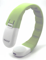 French bulldogs and a cavalier
King Charles spaniel. The other dogs were one each of an English bulldog
and a bullmastiff. A control group of 12 non-brachycephalic dogs were
included in the study, which lasted only one night for each dog at its
home. The neckband system reportedly is capable of measuring the
obstructive respiratory event index (OREI), which summarizes the rate of
obstructive SDB events per hour. The investigators report finding that
the BDs had a significantly higher OREI value and snore percentage than
the control dogs. They opined that the results would potentially be
different if the BD group consisted of more dogs with longer snouts such
as the bullmastiff and the cavalier. They found that the neckband system
was easy to use. They concluded that brachycephaly is associated with
SDB, and that the neckband system is a feasible way of characterizing
SDB in dogs.
French bulldogs and a cavalier
King Charles spaniel. The other dogs were one each of an English bulldog
and a bullmastiff. A control group of 12 non-brachycephalic dogs were
included in the study, which lasted only one night for each dog at its
home. The neckband system reportedly is capable of measuring the
obstructive respiratory event index (OREI), which summarizes the rate of
obstructive SDB events per hour. The investigators report finding that
the BDs had a significantly higher OREI value and snore percentage than
the control dogs. They opined that the results would potentially be
different if the BD group consisted of more dogs with longer snouts such
as the bullmastiff and the cavalier. They found that the neckband system
was easy to use. They concluded that brachycephaly is associated with
SDB, and that the neckband system is a feasible way of characterizing
SDB in dogs.
RETURN TO TOP
Breeders’ Responsibilities
BAOS is a consequence of the conformation standards for the CKCS. Cavaliers with significant breathing difficulties or that have required surgery to correct airway obstruction, should not be used for breeding.
 In
February 2023,
the OFA (Orthopedic Foundation of America)
joined the UK Kennel Club in examining dogs for symptoms of BOAS
(brachycephalic obstructive airway syndrome). The examination, called
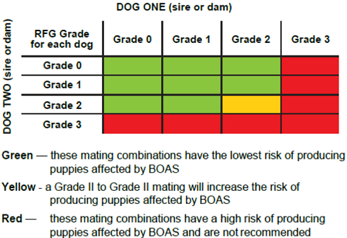
the Respiratory Function Grading Scheme, grades dogs on a scale from
Grade 0 to Grade III to objectively diagnose BOAS:
In
February 2023,
the OFA (Orthopedic Foundation of America)
joined the UK Kennel Club in examining dogs for symptoms of BOAS
(brachycephalic obstructive airway syndrome). The examination, called
the Respiratory Function Grading Scheme, grades dogs on a scale from
Grade 0 to Grade III to objectively diagnose BOAS:
• Grade 0 – the dog is clinically unaffected and free of any respiratory signs of BOAS (no evidence of disease, no BOAS related noise heard even with a stethoscope)
• Grade I – the dog is clinically unaffected but does have mild respiratory signs linked to BOAS (noise is mild and only audible with a stethoscope)
• Grade II – the dog is clinically affected and has moderate respiratory signs of BOAS (noise is audible even without a stethoscope)
• Grade III – the dog is clinically affected and has severe respiratory signs of BOAS (noise is audible even without a stethoscope)
Both Grade 0 and Grade I will be considered to be clinically normal and BOAS-unaffected as they exercise without difficulty and do not appear to have any clinical signs related to airway obstruction. These will receive OFA certification numbers, and their results will be posted on the OFA website. In Grades 2 and 3, when stertor or stridor noise is heard without a stethoscope, these dogs are considered BOAS-affected with clinical signs affecting quality of life. Results for dogs with Grade 2 or 3 results will only be posted on the OFA website if their owners authorize release of abnormal data. All results will be shared with an international team of collaborators for statistical purposes, but individual abnormal results will never be publicly released unless specifically authorized. For additional details, see the OFA announcement.
Using the RFGS grades and the guidelines in the chart below, breeders may reduce the chances of producing puppies affected by BOAS. However, since the inheritance of BOAS is not fully understood and is not entirely predictable, this guidance cannot guarantee that all puppies from unaffected parents will be free of BOAS.
RETURN TO TOP
Research News
February 2024:
Finnish study finds cavaliers rank high among breeds at risk for
sleep-disorderd breathing.
 In
a
February 2024 article, a team of Finnish researchers (Iida
Niinikoski [right], Sari-Leena Himanen, Mirja Tenhunen, Mimma
Aromaa, Liisa Lilja-Maula, Minna M. Rajamäki) evaluated the risk factors
for sleep-disordered breathing (SDB) in serveral dog breeds. Of 28
brachycephalic dogs, 8 (28.5%) were cavalier King Charles spaniels.
Other SDB breeds comprised 35 dogs. They reported that "We had an
overrepresentation of Cavalier King Charles Spaniels, French Bulldogs
and Labrador Retrievers." They found that brachycephaly was a
significant risk factor for SDB, particularly apnea, and BOAS-positive
class (moderate or severe BOAS signs) was a significant risk factor,
along with excess weight. Aging and Chiari-like malformation were not
risk factors among the dogs in this study.
In
a
February 2024 article, a team of Finnish researchers (Iida
Niinikoski [right], Sari-Leena Himanen, Mirja Tenhunen, Mimma
Aromaa, Liisa Lilja-Maula, Minna M. Rajamäki) evaluated the risk factors
for sleep-disordered breathing (SDB) in serveral dog breeds. Of 28
brachycephalic dogs, 8 (28.5%) were cavalier King Charles spaniels.
Other SDB breeds comprised 35 dogs. They reported that "We had an
overrepresentation of Cavalier King Charles Spaniels, French Bulldogs
and Labrador Retrievers." They found that brachycephaly was a
significant risk factor for SDB, particularly apnea, and BOAS-positive
class (moderate or severe BOAS signs) was a significant risk factor,
along with excess weight. Aging and Chiari-like malformation were not
risk factors among the dogs in this study.
June 2023:
Swedish study of 327 breeds finds cavaliers rank in top
five of high risk breeds for brachycephalic obstructive airway syndrome
disorders.
 In a
May 2023 article, Swedish researchers M. Dimopoulou
(right), K.
Engdah, J. Ladlow, G. Andersson, Å. Hedhammar, E. Skiöldebrand, and I.
Ljungvall examined the medical records from 2011 through 2014 of 450,000
dogs of 327 breeds, for upper respiratory tract disorders (URTs). They
included calculatations of the breed-specific incidence rate for each
breed. Their findings include:
In a
May 2023 article, Swedish researchers M. Dimopoulou
(right), K.
Engdah, J. Ladlow, G. Andersson, Å. Hedhammar, E. Skiöldebrand, and I.
Ljungvall examined the medical records from 2011 through 2014 of 450,000
dogs of 327 breeds, for upper respiratory tract disorders (URTs). They
included calculatations of the breed-specific incidence rate for each
breed. Their findings include:
• "The Boston terrier, boxer, CKCS [cavalier King Charles spaniel], English and French bulldog and pug are brachycephalic breeds and affected to a variable degree by BOAS [brachycephalic obstructive airway syndrome]."
• "Among 13 breeds with high RR [relative risk] for URT disorders in this study, eight (Boston terrier, boxer, CKCS, English bulldog, French bulldog, Japanese chin, Pomeranian, pug) are recognised as brachycephalic by most researchers."
• "The brachycephalic conformation has been linked to an increased risk for URT disorders, and specifically BOAS, in the French and English bulldog, the CKCS, Boston terrier and the pug."
• "The Chihuahua, CKCS, Pomeranian and Yorkshire terrier are breeds frequently affected with the URT disorder tracheal collapse."
• "The boxer, CKCS, Chihuahua, French bulldog, pug and standard poodle had high risk for co-morbidity both prior and after the first URT claim."
May 2023:
Finnish researchers use electronic neckband to track
sleep-disordered breathing in brachycephalic dogs.
In
a
May 2023 article, a team of Finnish researchers (Iida Niinikoski,
Sari-Leena Himanen, Mirja Tenhunen, Liisa Lilja-Maula, Minna M.
Rajamäki) evaluated the use of a portable neckband manufactured by
Nukute Ltd. for detecting sleep-disordered breathing (SDB) in 12
brachycephalic dogs (BDs), including 9 French bulldogs and a cavalier
King Charles spaniel. The other dogs were one each of an English bulldog
and a bullmastiff. A control group of 12 non-brachycephalic dogs were
included in the study, which lasted only one night for each dog at its
home. The neckband system reportedly is capable of measuring the
obstructive respiratory event index (OREI), which summarizes the rate of
obstructive SDB events per hour. The investigators report finding that
the BDs had a significantly higher OREI value and snore percentage than
the control dogs. They opined that the results would potentially be
different if the BD group consisted of more dogs with longer snouts such
as the bullmastiff and the cavalier. They found that the neckband system
was easy to use. They concluded that brachycephaly is associated with
SDB, and that the neckband system is a feasible way of characterizing
SDB in dogs.
April 2023:
Brachycephalic obstructive airway syndrome (BOAS) affected left
atrium parameters independent of mitral valve disease, in a Slovenia
study.
 In a
February 2023 article, Slovenian researchers (M. Brložnik, A. Nemec
Svete, V. Erjavec & A. Domanjko Petric [right]) compared the echocardiographic
measurements in three brachycephalic dog breeds -- 30 French bulldogs,
15 pugs, and 12 Boston terriers -- having signs of brachycephalic
obstructive airway syndrome (BOAS), to determine possible
echocardiographic differences between brachycephalic and
non-brachycephalic dogs and to evaluate possible echocardiographic
changes due to BOAS. There obtained several echocardiographic
measurements, including the left artium-to-aorta ratio (LA/Ao). They
report finding that the LA/Ao values for the French bulldogs were 1.56
(1.44-1.60), and for pugs were 1.49 (1.45-1.62), and for Boston terriers
were 1.55 (1.50-1.68). They found that for all three breeds, dogs with
signs of BAOS had different parameters than did dogs of the same breed
without BAOS, as well as significant differences in echocardiographic
parameters between the dogs of the three brachycephalic breeds and
non-brachycephalic dogs. They concluded:
In a
February 2023 article, Slovenian researchers (M. Brložnik, A. Nemec
Svete, V. Erjavec & A. Domanjko Petric [right]) compared the echocardiographic
measurements in three brachycephalic dog breeds -- 30 French bulldogs,
15 pugs, and 12 Boston terriers -- having signs of brachycephalic
obstructive airway syndrome (BOAS), to determine possible
echocardiographic differences between brachycephalic and
non-brachycephalic dogs and to evaluate possible echocardiographic
changes due to BOAS. There obtained several echocardiographic
measurements, including the left artium-to-aorta ratio (LA/Ao). They
report finding that the LA/Ao values for the French bulldogs were 1.56
(1.44-1.60), and for pugs were 1.49 (1.45-1.62), and for Boston terriers
were 1.55 (1.50-1.68). They found that for all three breeds, dogs with
signs of BAOS had different parameters than did dogs of the same breed
without BAOS, as well as significant differences in echocardiographic
parameters between the dogs of the three brachycephalic breeds and
non-brachycephalic dogs. They concluded:
"We found significant differences in echocardiographic parameters between dogs of the three brachycephalic breeds and non-brachycephalic dogs, implying that breed-specific echocardiographic reference values should be used in clinical practice. In addition, significant differences were observed between brachycephalic dogs with and without signs of BOAS. The observed echocardiographic differences suggest higher right heart diastolic pressures affecting right heart function in brachycephalic dogs with and without signs of BOAS, and several of the differences are consistent with findings in OSA patients. Most of the changes of the heart morphology and function can be attributed to brachycephaly alone and not to the symptomatic stage."
 EDITOR'S
NOTE: We find from this study that dogs with BAOS can have
changes in their hearts' measurements and functions due not to
mitral valve disease (MVD) at all, but due to the BAOS. This means
that cardiologists cannot continue to rely upon simplistic species-wide
echocardiographic measurements to diagnose the presence of heart
enlargement, or even the existence of MVD at all, in brachycephalic dogs
if they have symptoms of BAOS. Cardiologists now are faced with a new
learning curve.
EDITOR'S
NOTE: We find from this study that dogs with BAOS can have
changes in their hearts' measurements and functions due not to
mitral valve disease (MVD) at all, but due to the BAOS. This means
that cardiologists cannot continue to rely upon simplistic species-wide
echocardiographic measurements to diagnose the presence of heart
enlargement, or even the existence of MVD at all, in brachycephalic dogs
if they have symptoms of BAOS. Cardiologists now are faced with a new
learning curve.
February 2023:
OFA announces the Respiratory Function Grading Scheme testing
for BOAS. OFA (Orthopedic Foundation of America) has
joined the UK Kennel Club in examining dogs for symptoms of BOAS
(brachycephalic obstructive airway syndrome). The examination, called
the Respiratory Function Grading Scheme, grades dogs on a scale from
Grade 0 to Grade III to objectively diagnose BOAS:
• Grade 0 – the dog is clinically unaffected and free of any respiratory signs of BOAS (no evidence of disease, no BOAS related noise heard even with a stethoscope)
• Grade I – the dog is clinically unaffected but does have mild respiratory signs linked to BOAS (noise is mild and only audible with a stethoscope)
• Grade II – the dog is clinically affected and has moderate respiratory signs of BOAS (noise is audible even without a stethoscope)
• Grade III – the dog is clinically affected and has severe respiratory signs of BOAS (noise is audible even without a stethoscope)
Both Grades 0 and I will be considered to be clinically normal and BOAS-unaffected as they exercise without difficulty and do not appear to have any clinical signs related to airway obstruction. These will receive OFA certification numbers, and their results will be posted on the OFA website. In Grades 2 and 3, when stertor or stridor noise is heard without a stethoscope, these dogs are considered BOAS-affected with clinical signs affecting quality of life. Results for dogs with Grade 2 or 3 results will only be posted on the OFA website if their owners authorize release of abnormal data. All results will be shared with an international team of collaborators for statistical purposes, but individual abnormal results will never be publicly released unless specifically authorized. For additional details, see the OFA announcement.
April 2021:
Cavalier with sleep disordered breathing (SDB) is successfully
fitted with a ventilation mask every night.
 In an
April 2021 article, a team of Australian veterinary specialists
(Waylon Wiseman [right], A. Rosenblatt, M. Kunga)
report a case of a 4-year-old female cavalier King Charles spaniel
experiencing sleep apnea episodes every 10 to 15 minutes during sleep.
They diagnosed "dynamic pharyngeal collapse" (DPC), which involves
partial or complete collapse of the pharynx. They performed a series of
surgical procedures -- bilateral tonsillectomy, bilateral laryngeal
sacculectomy, and unilateral cricoarytenoid lateralisation -- with
minimal improvement. The cavalier's breathing pattern quality improved
while awake, but she continued to suffer from SBD at night, with apnea
episodes every 30 minutes. The dog was fitted with a "continuous
positive airway pressure" (CPAP) device, which included a mask connected
to an air hose and pump, to create a mild positive pressure in the dog's
airway. (See Figures 1 & 2.) A muzzle was fitted over the mask to secure
it to the dog's head. The cavalier gradually got accustomed to the
device, with the owner using positive reinforcement with treats and
removing the apparatus before the dog displayed signs of distress.
Eventually, after continuing to suffer SDB episodes without the device,
the dog sought out the mask and muzzle. With the CPAP device in place,
the cavalier was able to sleep between 4 to 5 hours each night. Four
years later, when the cavalier was 8 years old, she was examined with a
dynamic fluoroscope to determine if the SDB was due to DPC. The
fluoroscopy exam confirmed partial pharyngeal collapse.
In an
April 2021 article, a team of Australian veterinary specialists
(Waylon Wiseman [right], A. Rosenblatt, M. Kunga)
report a case of a 4-year-old female cavalier King Charles spaniel
experiencing sleep apnea episodes every 10 to 15 minutes during sleep.
They diagnosed "dynamic pharyngeal collapse" (DPC), which involves
partial or complete collapse of the pharynx. They performed a series of
surgical procedures -- bilateral tonsillectomy, bilateral laryngeal
sacculectomy, and unilateral cricoarytenoid lateralisation -- with
minimal improvement. The cavalier's breathing pattern quality improved
while awake, but she continued to suffer from SBD at night, with apnea
episodes every 30 minutes. The dog was fitted with a "continuous
positive airway pressure" (CPAP) device, which included a mask connected
to an air hose and pump, to create a mild positive pressure in the dog's
airway. (See Figures 1 & 2.) A muzzle was fitted over the mask to secure
it to the dog's head. The cavalier gradually got accustomed to the
device, with the owner using positive reinforcement with treats and
removing the apparatus before the dog displayed signs of distress.
Eventually, after continuing to suffer SDB episodes without the device,
the dog sought out the mask and muzzle. With the CPAP device in place,
the cavalier was able to sleep between 4 to 5 hours each night. Four
years later, when the cavalier was 8 years old, she was examined with a
dynamic fluoroscope to determine if the SDB was due to DPC. The
fluoroscopy exam confirmed partial pharyngeal collapse.
February 2021:
Drs. Rusbridge and Knowler find impaired cerebrospinal fluid
circulation predisposes Chiari-like malformation and syringomyelia in
cavaliers.
 In
a
February 2021 article, neurology researchers Clare Rusbridge and
Penny Knowler [right] review current data linking cerebrospinal
fluid (CSF) disorders with brachyephaly and shorten muzzles in cavalier
King Charles spaniels and other small breed dogs. They point out that
there is increasing evidence that brachycephaly disrupts CSF movement
and absorption, predisposing ventriculomegaly, hydrocephalus, and
quadrigeminal cistern expansion, as well as Chiari-like malformation and
syringomyelia. They show how the reduction of the lymphatic absorption
of CSF through the lymphatic system organs located in the nasal and
skull base, combined with the restriction of CSF movement through the
junction of the skull with the spinal cord, appear to be key
consequences of extreme brachycephaly in dogs and explain the likely
causes of these neurological disorders..
In
a
February 2021 article, neurology researchers Clare Rusbridge and
Penny Knowler [right] review current data linking cerebrospinal
fluid (CSF) disorders with brachyephaly and shorten muzzles in cavalier
King Charles spaniels and other small breed dogs. They point out that
there is increasing evidence that brachycephaly disrupts CSF movement
and absorption, predisposing ventriculomegaly, hydrocephalus, and
quadrigeminal cistern expansion, as well as Chiari-like malformation and
syringomyelia. They show how the reduction of the lymphatic absorption
of CSF through the lymphatic system organs located in the nasal and
skull base, combined with the restriction of CSF movement through the
junction of the skull with the spinal cord, appear to be key
consequences of extreme brachycephaly in dogs and explain the likely
causes of these neurological disorders..
October 2020:
Cavaliers ranked third among brachycephalic breeds requiring
veterinary care in the UK in 2016.
 In an
October 2020 article by a team of Royal Veterinary College
researchers (D. G. O’Neill [right], C. Pegram, P. Crocker, D.
C. Brodbelt, D. B. Church, R. M. A. Packer), they examined the 2016
primary-care veterinary records of a random sample of 22,333 dogs to
identify predispositions and protections in brachycephalic dogs. Of
those dogs, 4,169 (18.74%) were of brachycephalic breeds and the
remaining 18,079 (81.26%) were non-brachycephalic. The most common
brachycephalic breeds were Chihuahuas (955 dogs, 22.91%), Shih-tzus (795
dogs, 19.07%) and cavalier King Charles spaniels (435 dogs, 10.43%). The
most common disorders (i.e. greatest prevalence) which they found in the
brachycephalic types were periodontal disease, otitis externa, obesity,
anal sac impaction, overgrown nail(s), diarrhea, and heart murmur. Two
disorders which had reduced odds for brachycephalic types were
undesirable behavior and claw injury. Regarding the CKCS specifically,
they report that:
In an
October 2020 article by a team of Royal Veterinary College
researchers (D. G. O’Neill [right], C. Pegram, P. Crocker, D.
C. Brodbelt, D. B. Church, R. M. A. Packer), they examined the 2016
primary-care veterinary records of a random sample of 22,333 dogs to
identify predispositions and protections in brachycephalic dogs. Of
those dogs, 4,169 (18.74%) were of brachycephalic breeds and the
remaining 18,079 (81.26%) were non-brachycephalic. The most common
brachycephalic breeds were Chihuahuas (955 dogs, 22.91%), Shih-tzus (795
dogs, 19.07%) and cavalier King Charles spaniels (435 dogs, 10.43%). The
most common disorders (i.e. greatest prevalence) which they found in the
brachycephalic types were periodontal disease, otitis externa, obesity,
anal sac impaction, overgrown nail(s), diarrhea, and heart murmur. Two
disorders which had reduced odds for brachycephalic types were
undesirable behavior and claw injury. Regarding the CKCS specifically,
they report that:
"The degree (or severity) of brachycephaly varies between breeds (a bulldog may be considered as more severely brachycephalic than a Cavalier King Charles Spaniel) but there can also be considerable variation in brachycephaly within breeds. Shifting the median severity of brachycephaly towards a longer skull shape within breeds has been suggested as one option to reduce the prevalence of disorders directly linked to brachycephaly while still retaining these breeds within the overall dog population."
They concluded that:
"This study provides strong evidence to support the common assertion that brachycephalic breeds are generally less healthy than their non-brachycephalic counterparts in relation to total disorder counts and specific common conditions recorded. Potential solutions to some of these health problems are likely to require conformational change to current skull shapes averages for many breeds; however, many other health problems will require targeted action at the individual breed level, owing to large differences in individual breed predispositions to disorders."
May 2020:
German study of cavalier nasal passages shows rapid elongation
of facial shape has created the breed's complex nasal skeleton.
 In
a
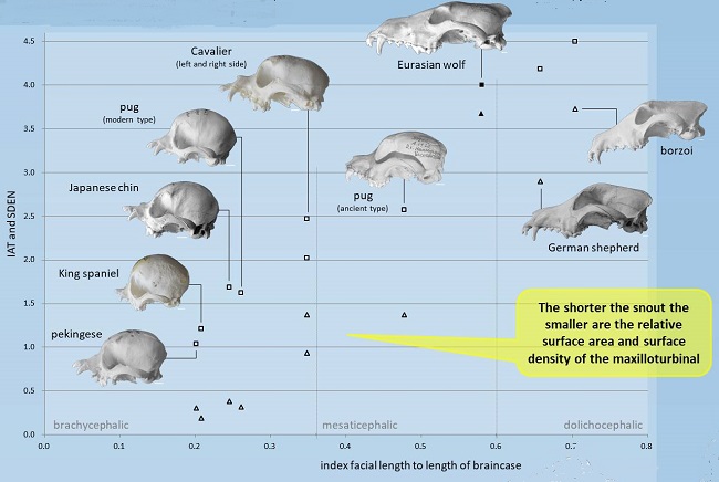
May 2020 article, two mammalogists, Franziska Wagner and Irina Ruf
(right), have examined the skulls of five toy dog breeds
(Japanese chin, King Charles spaniel, cavalier King Charles spaniel,
pekingese, and pug), to compare the consequences of breeders'
intentional genetic mutations upon the nasal structures -- particularly
the turbinates (air-filters) of the resulting generations of the breeds.
The investigators found that because the cavalier has resulted from a
relatively rapid (since the year 1928) elongation of its snout from its
King Charles spaniel ancestor (called "reverse breeding"), its turbinal
skeleton has changed very little from that of its ancestors. As for
other breeds, they found that with decreasing snout length, the nasal
cavity is minimized and the seven turbinals suffer as a consequence,
especially the interturbinals, which are reduced in number during their
developmental period.
In
a
May 2020 article, two mammalogists, Franziska Wagner and Irina Ruf
(right), have examined the skulls of five toy dog breeds
(Japanese chin, King Charles spaniel, cavalier King Charles spaniel,
pekingese, and pug), to compare the consequences of breeders'
intentional genetic mutations upon the nasal structures -- particularly
the turbinates (air-filters) of the resulting generations of the breeds.
The investigators found that because the cavalier has resulted from a
relatively rapid (since the year 1928) elongation of its snout from its
King Charles spaniel ancestor (called "reverse breeding"), its turbinal
skeleton has changed very little from that of its ancestors. As for
other breeds, they found that with decreasing snout length, the nasal
cavity is minimized and the seven turbinals suffer as a consequence,
especially the interturbinals, which are reduced in number during their
developmental period.
February 2020:
Australian study finds cavaliers ranked as second highest breed
for brachycephalic airway surgeries.
 In
a
February 2020 article, researchers (Barbara Lindsay [right],
D Cook, J-M Wetzel, S Siess, P Moses) examined the medical records of
248 dogs which were surgically treated for brachycephalic conditions
from 2001 through 2012 in Australian veterinary specialty hospitals. Of
those, 42 (16.9%) were cavalier King Charles spaniels. The only breed
with more cases was the pug with 44 (17.7%) cases. The third highest
breed was the English bulldog with 31 (12.5%) cases. The most common
disorder which was surgically treated was elongated soft palate in 242
(97.6%) dogs. Others were hyperplastic tonsillar tissue (106 dogs --
42.7%), everted saccules (103 dogs -- 41.5%), and stenotic nares (97 dos
-- 39.1%). The report details different complications experienced by the
dogs prior to, during, and following surgery, but does not categorize
them by breed. Since 97.6% of the surgeries were for elongated soft
palates, and since that disorder is the most common one among cavaliers,
it may be assumed that palate surgery was the main one for the CKCSs in
this study.
In
a
February 2020 article, researchers (Barbara Lindsay [right],
D Cook, J-M Wetzel, S Siess, P Moses) examined the medical records of
248 dogs which were surgically treated for brachycephalic conditions
from 2001 through 2012 in Australian veterinary specialty hospitals. Of
those, 42 (16.9%) were cavalier King Charles spaniels. The only breed
with more cases was the pug with 44 (17.7%) cases. The third highest
breed was the English bulldog with 31 (12.5%) cases. The most common
disorder which was surgically treated was elongated soft palate in 242
(97.6%) dogs. Others were hyperplastic tonsillar tissue (106 dogs --
42.7%), everted saccules (103 dogs -- 41.5%), and stenotic nares (97 dos
-- 39.1%). The report details different complications experienced by the
dogs prior to, during, and following surgery, but does not categorize
them by breed. Since 97.6% of the surgeries were for elongated soft
palates, and since that disorder is the most common one among cavaliers,
it may be assumed that palate surgery was the main one for the CKCSs in
this study.
December 2018:
Australian
veterinarians urge that cavaliers not be bred due to genetic
brachycephalic conditions.
 In a
January 2019 article, a team of
Australian veterinarians at the University of Sydney, led by ethicist
Anne Fawcett (right) reviews several brachycephalic conditions in
cavalier King Charles spaniels (CKCS) and other breeds (Affenpinscher,
American bulldog, Australian bulldog, Australian bulldog miniature,
Boston terrier, boxer, British bulldog, Dogue De Bordeaux, French
bulldog, Griffon, Griffon Brabancon, Griffon Bruxellois, Lhasa Apso,
mastiff, Neopolitan mastiff, Pekingese, pug and Shih Tzu). In
particular, they attribute primary secretory otitis media (PSOM),
Chiari-like malformation (CM), syringomyelia (SM), and
patellar luxation
in the cavalier upon brachycephalic airway obstruction syndrome (BAOS).
They conclude by stating:
In a
January 2019 article, a team of
Australian veterinarians at the University of Sydney, led by ethicist
Anne Fawcett (right) reviews several brachycephalic conditions in
cavalier King Charles spaniels (CKCS) and other breeds (Affenpinscher,
American bulldog, Australian bulldog, Australian bulldog miniature,
Boston terrier, boxer, British bulldog, Dogue De Bordeaux, French
bulldog, Griffon, Griffon Brabancon, Griffon Bruxellois, Lhasa Apso,
mastiff, Neopolitan mastiff, Pekingese, pug and Shih Tzu). In
particular, they attribute primary secretory otitis media (PSOM),
Chiari-like malformation (CM), syringomyelia (SM), and
patellar luxation
in the cavalier upon brachycephalic airway obstruction syndrome (BAOS).
They conclude by stating:
"In summary, dogs with BOAS do not enjoy freedom from discomfort, nor freedom from pain, injury, and disease, and they do not enjoy the freedom to express normal behaviour. According to both deontological and utilitarian ethical frameworks, the breeding of dogs with BOAS cannot be justified, and further, cannot be recommended, and indeed, should be discouraged by veterinarians."
September 2018:
UK brachycephaly surgeons treat five cavaliers for sleep apnea.
 In
an
April 2018 abstract, a team of UK veterinary surgeons specializing
in treating brachycephalic animals (Tom Hinchliffe, Nai-Chieh Liu, Jane
Ladlow [right]) report their
study of five cavalier King Charles spaniels which had been suffering
from obstructive sleep apnea (apnoea). Apnea means the suspension of
breathing due to blocked airways. Previously, it had been reported only
in English bulldogs, French bulldogs, and pugs, all typically severely
brachycephalic breeds. All five CKSCs suffered from heavy snoring and
noisy breathing and choking, as well as sleep apnea. In addition, all
five had eosinophilic stomatitis and primary secretory otitis media
(PSOM); four had mitral valve disease (MVD); and three had syringomyelia
(SM). Also, all five had "aberrant nasal turbinates, nasal septal
deviation and soft palate thickening and 3/5 had nasopharyngeal
thickening and tracheal collapse."
In
an
April 2018 abstract, a team of UK veterinary surgeons specializing
in treating brachycephalic animals (Tom Hinchliffe, Nai-Chieh Liu, Jane
Ladlow [right]) report their
study of five cavalier King Charles spaniels which had been suffering
from obstructive sleep apnea (apnoea). Apnea means the suspension of
breathing due to blocked airways. Previously, it had been reported only
in English bulldogs, French bulldogs, and pugs, all typically severely
brachycephalic breeds. All five CKSCs suffered from heavy snoring and
noisy breathing and choking, as well as sleep apnea. In addition, all
five had eosinophilic stomatitis and primary secretory otitis media
(PSOM); four had mitral valve disease (MVD); and three had syringomyelia
(SM). Also, all five had "aberrant nasal turbinates, nasal septal
deviation and soft palate thickening and 3/5 had nasopharyngeal
thickening and tracheal collapse."
Surgical treatment consisted of a combination of laser turbinectomy, folding flap palatoplasty, tonsillectomy, laryngeal sacculectomy and cuneiform process resection. All cases improved within a week, and four of them were resolved completely. (See also this December 2018 article on the same study.)
June 2018:
Italian veterinarians report 3 surgeries on a cavalier to
relieve severe laryngeal collapse.
 In
a
June 2018 article about permanent tracheostomies performed on 15
brachycephalic dogs with severe laryngeal collapse, a team of Italian
veterinary surgeons (Matteo Gobbetti [right], Stefano Romussi,
Paolo Buracco, Valerio Bronzo, Samuele Gatti, Matteo Cantatore) reported
on three surgeries required to resolve grade 3 laryngeal collapse in a
cavalier King Charles spaniel. The CKCS was an 11 year old female. The
first procedure shorted the dog's elongated soft palate. A month later,
the surgeons resected the cavalier's hypertrophic aryepiglottic folds
surrounding the larynx. A month after the second procedure, the surgeons
performed a permanent tracheostomy, which creates a permanent opening (a
stoma) through the neck into the trachea (windpipe). A tracheostomy tube
or trach tube is placed through the stoma to provide an airway and to
remove secretions from the lungs. The CKCS survived the three surgeries
successfully and lived another 143 days, dying by euthanasia due to a
separate medical disorder.
In
a
June 2018 article about permanent tracheostomies performed on 15
brachycephalic dogs with severe laryngeal collapse, a team of Italian
veterinary surgeons (Matteo Gobbetti [right], Stefano Romussi,
Paolo Buracco, Valerio Bronzo, Samuele Gatti, Matteo Cantatore) reported
on three surgeries required to resolve grade 3 laryngeal collapse in a
cavalier King Charles spaniel. The CKCS was an 11 year old female. The
first procedure shorted the dog's elongated soft palate. A month later,
the surgeons resected the cavalier's hypertrophic aryepiglottic folds
surrounding the larynx. A month after the second procedure, the surgeons
performed a permanent tracheostomy, which creates a permanent opening (a
stoma) through the neck into the trachea (windpipe). A tracheostomy tube
or trach tube is placed through the stoma to provide an airway and to
remove secretions from the lungs. The CKCS survived the three surgeries
successfully and lived another 143 days, dying by euthanasia due to a
separate medical disorder.
Overall among the 15 dogs, major complications were diagnosed in 12 (80%) dogs, resulting in death in 8 and revision surgery in 4 dogs, including the CKCS. They report finding that permanent tracheostomy was associated with a high risk of complications and postoperative death in brachycephalic dogs. However, long-term survival (exceeding 5 years) with a good quality of life was documented in 5 of 15 dogs, including the CKCS. They concluded that "permanent tracheostomy is a suitable salvage option in brachycephalic dogs with severe laryngeal collapse that did not improve following more conservative surgeries."
January 2018:
UK study finds MRI images can assess oral cavity landmarks and
that soft-palate-to-epiglottis overlap is directly proportionate to
degree of brachcephalia in dogs.
 In
a
February 2018 article, a team of UK researchers (David A. Barker
[right], Carlos Rubiños, Olivier Taeymans, Jackie L. Demetriou)
used magnetic resonance imaging (MRI) to evaluate the anatomy of the
oral cavity of 50 brachycephalic dogs compared to 50 nonbrachycephalic
dogs. Five of the brachycephalic dogs were cavalier King Charles
spaniels. The researchers report that MRI scans are effective to assess
anatomic landmarks and thicknesses in the dogs' oral cavities. They
found that the brachycephalic dogs had significantly thicker soft
palates, and that the degree of overlap of the soft palate and the
epiglottis (the flap of elastic cartilage at the root of the tongue,
attached to the entrance of the larynx) was significantly greater in
brachycephalic dogs and directly proportional to the degree of
brachycephalia.
In
a
February 2018 article, a team of UK researchers (David A. Barker
[right], Carlos Rubiños, Olivier Taeymans, Jackie L. Demetriou)
used magnetic resonance imaging (MRI) to evaluate the anatomy of the
oral cavity of 50 brachycephalic dogs compared to 50 nonbrachycephalic
dogs. Five of the brachycephalic dogs were cavalier King Charles
spaniels. The researchers report that MRI scans are effective to assess
anatomic landmarks and thicknesses in the dogs' oral cavities. They
found that the brachycephalic dogs had significantly thicker soft
palates, and that the degree of overlap of the soft palate and the
epiglottis (the flap of elastic cartilage at the root of the tongue,
attached to the entrance of the larynx) was significantly greater in
brachycephalic dogs and directly proportional to the degree of
brachycephalia.
May 2017:
Nationwide insurance reports cavaliers and other
brachycephalic breeds are more often affected by common health
conditions.
 In a
March 2017 report, Nationwide Insurance reviews its
database of claims made for more than 1.27 million dogs of 300+ breeds
and mixes, from both
brachycephalic and non-brachycephalic breeds, over a nine-year period
(2007-2015). Cavalier King Charles spaniels were included among the 24
brachycephalic breeds. The report shows that, after excluding typically
brachycephalic conditions (e.g., stenotic nares, elongated
soft palate, everted laryngeal saccules, tracheal hypoplasia or
stenosis), brachy dogs are less healthy than dogs with a more normal
canine appearance. Specifically, Nationwide reports higher prevalences
of:
In a
March 2017 report, Nationwide Insurance reviews its
database of claims made for more than 1.27 million dogs of 300+ breeds
and mixes, from both
brachycephalic and non-brachycephalic breeds, over a nine-year period
(2007-2015). Cavalier King Charles spaniels were included among the 24
brachycephalic breeds. The report shows that, after excluding typically
brachycephalic conditions (e.g., stenotic nares, elongated
soft palate, everted laryngeal saccules, tracheal hypoplasia or
stenosis), brachy dogs are less healthy than dogs with a more normal
canine appearance. Specifically, Nationwide reports higher prevalences
of:
• Dermatological diseases
• Gastrointestinal tract issues
• Poorly aligned dentition and tooth extractions
• Breathing issues (heatstroke, hyperthermia, pneumonia)
• Heart disease
• Orthopedic and spinal issues (patellar luxation, intervertebral disc disease)
January 2017:
UK study identifies characteristics that cause painful
Chiari-like malformation and syringomyelia in cavaliers.
In
a
January 2017 article, a team of veteran neurologists and other
specialists (Susan P. Knowler [right], Chloe Cross, Sandra
Griffiths, Angus K. McFadyen, Jelena Jovanovik, Anna Tauro, Zoha Kibar,
Colin J. Driver, Roberto M. La Ragione, Clare Rusbridge) used novel
brain-mapping techniques to compare distance and angle measurements from
MRI scans of 162 cavalier King Charles spaniels to thereby determine the
relationship between the measure of physical characteristics obtainable
from MRI scans and resulting risk of pain caused by Chiari-like
malformation (CM) and syringomyelia (SM). They found that CM pain
appears to be associated with increased brachycephaly (shorteness of
muzzle), and that SM can result from different combinations of
abnormalities of the forebrain, caudal fossa, and craniocervical
junction which make up the neural parenchyma, and impede the flow of
cerebrospinal fluid.
The study includes
a motion picture which highlights the dynamic changes of the skull
conformation and brain parenchyma associated with progressive
brachycephaly and airorhynchy [the upward rotation of the front of the
palate]. As the video morphs from the control dog to one with CM pain
that the nasal bone and hard palate become closer so that the hard
palate becomes more horizontal, the nasal cavity and frontal cavity
reduce in
 volume
and the rostral forebrain is flattened. As the model progresses into SM
case 1 the nasal and rostral forebrain changes become more extreme. In
addition to the forebrain changes, the hindbrain is pushed down and the
craniocervical junction kinks as a consequence of the cervical vertebral
being closer to the skull with flattening of the supraoccipital bone.
Consequently there is a “concertina” flexure of the brain with a
compensatory increase in height of the cranial fossa. In SM case 2 the
concertina flexure is more extreme lower with the cerebellum appear to
be folded back under the occipital lobe, and the olfactory bulbs are
much in size and displaced.
volume
and the rostral forebrain is flattened. As the model progresses into SM
case 1 the nasal and rostral forebrain changes become more extreme. In
addition to the forebrain changes, the hindbrain is pushed down and the
craniocervical junction kinks as a consequence of the cervical vertebral
being closer to the skull with flattening of the supraoccipital bone.
Consequently there is a “concertina” flexure of the brain with a
compensatory increase in height of the cranial fossa. In SM case 2 the
concertina flexure is more extreme lower with the cerebellum appear to
be folded back under the occipital lobe, and the olfactory bulbs are
much in size and displaced.
August 2015: UK researchers opine that corneal ulcers in cavaliers may be due to the breed standard favoring large eyes. In a May 2015 study by a team of researchers (Rowena M. A. Packer, Anke Hendricks, Charlotte C. Burn) from the UK's Royal Veterinary College, they measured eleven conformational features demonstrated to be breed-defining (muzzle length, cranial length, head width, eye width, neck length, neck girth, chest girth, chest width, body length, height at the withers and height at the base of tail) of 700 dogs, 31 dogs of which were affected with corneal ulcers, including three cavalier King Charles spaniels. They specifically criticized the CKCS breed standard for considering "large" eyes as a desirable feature, and also noted that the cavalier's predisposition to dry eye can lead to corneal ulcers. They stated:
"Several brachycephalic breeds have been identified as being predisposed to dry eye, including the Bulldog, Lhasa Apso, Shih Tzu, Pug, Pekingese, Boston Terrier and Cavalier King Charles Spaniel. Even moderately lowered tear production associated with dry eye may produce clinical signs in brachycephalic dogs, as a larger portion of the globe is exposed. In a UK based study, a higher proportion of brachycephalic dogs that were affected by dry eye were also affected by ulcers, than were non-brachycephalic dogs with dry eye, e.g. 36% of Shih Tzus and 30% of Cavalier King Charles Spaniels versus 17% of dogs in the overall study population."
They devised morphometric predictors for corneal ulcers, including
the "craniofacial ratio" (CFR), "the
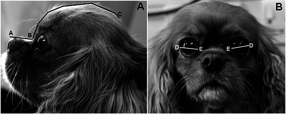 muzzle
length divided by the cranial length, which quantifies the degree of
brachycephaly", to differentiate skull morphologies. [Photos at
right: "This Cavalier King Charles Spaniel has a craniofacial ratio of
0.27 (muzzle length 28mm / cranial length 102mm), and a relative
palpebral fissure width value of 33.3% ((palpebral fissure width 34mm /
cranial length 102mm) *100"]
muzzle
length divided by the cranial length, which quantifies the degree of
brachycephaly", to differentiate skull morphologies. [Photos at
right: "This Cavalier King Charles Spaniel has a craniofacial ratio of
0.27 (muzzle length 28mm / cranial length 102mm), and a relative
palpebral fissure width value of 33.3% ((palpebral fissure width 34mm /
cranial length 102mm) *100"]
They found that brachycephalic dogs (craniofacial ratio <0.5) were twenty times more likely to have corneal ulcers than non-brachycephalic dogs. In addition, they found that: (a) a 10% increase in relative eyelid aperture width more than tripled the ulcer risk, and (b) exposed eye-white was associated with a nearly three times increased risk. They concluded that:
"The results demonstrate that artificially selecting for these facial characteristics greatly heightens the risk of corneal ulcers, and such selection should thus be discouraged to improve canine welfare."
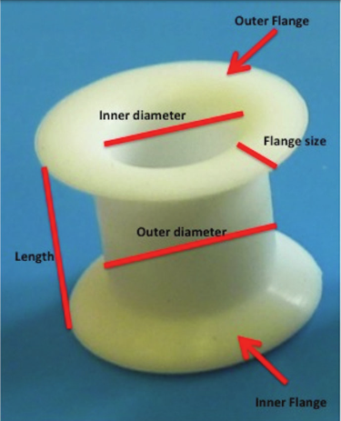 November 2014:
UK surgeons report limited success inserting tracheal stents for
temporary tracheostomies. In a
November 2014 study by a
group of UK veterinary surgeons (T. Trinterud, P. Nelissen, R. A. S.
White at Dick White Referrals), they report on the results of inserting
silicone tracheal stoma stents (right) for temporary maintenance of a
tracheostomy stoma for periods ranging from three hours to eight months,
in eighteen brachycephalic dogs, including three cavaliers. One of the
CKCSs was euthanized during its procedure and the stents in the other
two cavaliers had to be removed before the dogs were discharged from the
hospital.
November 2014:
UK surgeons report limited success inserting tracheal stents for
temporary tracheostomies. In a
November 2014 study by a
group of UK veterinary surgeons (T. Trinterud, P. Nelissen, R. A. S.
White at Dick White Referrals), they report on the results of inserting
silicone tracheal stoma stents (right) for temporary maintenance of a
tracheostomy stoma for periods ranging from three hours to eight months,
in eighteen brachycephalic dogs, including three cavaliers. One of the
CKCSs was euthanized during its procedure and the stents in the other
two cavaliers had to be removed before the dogs were discharged from the
hospital.
July 2014:
UK's Royal Veterinary College opens a clinic especially for
brachycephalic dogs.
![]() The RVC announced on July 1:
The RVC announced on July 1:
"A specialist clinic for brachycephalic dog breeds – also known as short-muzzled dogs – such as pugs, English and French bulldogs, cavalier King Charles spaniels and Pekingese, has been opened by the RVC at its Queen Mother Hospital for Animals in Hertfordshire.
"The Brachycephaly Clinic opened on Tuesday 1 July and is the first of its kind in the country exclusively specialising in the health of short-nosed dog breeds. This type of breed is one of the most popular pet choices in the UK, but the breeding of brachycephalic dogs has lead to a variety of health issues for the animals. These include problems with their bones and gait as well as eye, heart, ear (including hearing), skin, and breathing complications."
August 2013:
Canadian vet urges "Stop brachycephalism, now!", including cavaliers.
In a
 reasoned rant in a February 2013
Canadian Veterinary Journal article, veterinary dentist
Dr. Fraser
Hale (right) expresses his frustration with the mouths and teeth of
brachycephalic breeds, including the cavalier King Charles spaniel. In his
article, he states:
reasoned rant in a February 2013
Canadian Veterinary Journal article, veterinary dentist
Dr. Fraser
Hale (right) expresses his frustration with the mouths and teeth of
brachycephalic breeds, including the cavalier King Charles spaniel. In his
article, he states:
"In many Canadian jurisdictions, veterinarians have advocated for and achieved a ban on tail-docking, ear-cropping, and dewclaw removal as these are considered unnecessary cosmetic procedures that cause (temporary) pain with no benefit to the animals. I believe that as protectors of animal welfare, veterinarians should start a public awareness campaign to inform people of the serious, life-long negative impacts of brachycephalism. I believe we must stop referring to these conditions as 'normal for the breed' and refer to them as 'grossly abnormal in accordance with breed standards' because there is nothing remotely normal or desirable from the animal’s perspective. I believe we must stop using photographs of these deformed but comical breeds in advertising and promotional materials as this just increases public demand because they are 'so cute.'"
While he does not mention specific breeds in his article, in a subsequent interview, he stated:
"Of course, the small brachycephalics are the worst of the bunch. Oh yes, add CKCSs to the list."
 September 2012: UK
Dr. Gert ter Haar heads RVC's new ear, nose, and throat clinic.
UK's Royal Veterinary College has commenced an ear, nose, and throat referral
clinic at the Queen Mother Hospital for Animals in London. Dr. Gert ter
Haar (right) heads the unit, which offers services including diagnostics and
treatment of cavaliers with brachycephalic airway obstruction syndrome (BAOS),
advanced diagnostics and treatment for PSOM in cavaliers, as well as CT and BAER
diagnostics for deafness (plus hearing aid implants for selected patients).
Telephone 01707 666 365 for more information.
September 2012: UK
Dr. Gert ter Haar heads RVC's new ear, nose, and throat clinic.
UK's Royal Veterinary College has commenced an ear, nose, and throat referral
clinic at the Queen Mother Hospital for Animals in London. Dr. Gert ter
Haar (right) heads the unit, which offers services including diagnostics and
treatment of cavaliers with brachycephalic airway obstruction syndrome (BAOS),
advanced diagnostics and treatment for PSOM in cavaliers, as well as CT and BAER
diagnostics for deafness (plus hearing aid implants for selected patients).
Telephone 01707 666 365 for more information.
August 2012: US and Canadian researchers find that a mutation of the gene BMP3 plays a role in brachycephalic breeds. See the report here. Cavalier King Charles spaniels were included in the brachycephalic class of dogs studied.
March 2012: Cavaliers are listed among brachycephalic breeds requiring extra care when being anesthetized. In a March 2012 report by a team of Tufts University veterinary anesthesiologists, cavaliers are among brachycephalic breeds which require special attention when being sedated and anesthetized. Their advice includes: "Avoid excessive sedation. Avoid α2-agonists. Administer acepromazine at half dose. Preoxygenate. Use short-acting induction agent. Use appropriately sized endotracheal tubes. Extubate after patient is sitting up, vigorously chewing, bright, alert."
December 2010: Cavaliers were most common breed for BAOS surgery among 155 Australian dogs. In a study of BAOS surgery on 155 Australian dogs, the cavalier was the most common breed (29 dogs, 18.7%). The researchers found: "All CKCS had an elongated soft palate and accounted for 41% of the laryngeal collapse cases."
 October
2010: UK surgeon opines CKCS's brachycephalic disorders
and PSOM are related to Chiari-like malformation. Robert N.
White (right), a board certified veterinary soft tissue surgeon
practicing at Willows Veterinary Centre and Referral Service in
Solihull, West Midlands, observed in an
October 2010 presentation before a meeting of the UK's Association
of Veterinary Soft Tissue Surgeons, that the cavalier King Charles
spaniel does not appear to be a classically brachycephalic breed,
despite the extent of BAOS in the cavalier, and that the extent of both
PSOM and SM in the breed suggests that the CKCS may suffer from a
combination syndrome of the three disorders, all associated with
Chiari-like malformation.
October
2010: UK surgeon opines CKCS's brachycephalic disorders
and PSOM are related to Chiari-like malformation. Robert N.
White (right), a board certified veterinary soft tissue surgeon
practicing at Willows Veterinary Centre and Referral Service in
Solihull, West Midlands, observed in an
October 2010 presentation before a meeting of the UK's Association
of Veterinary Soft Tissue Surgeons, that the cavalier King Charles
spaniel does not appear to be a classically brachycephalic breed,
despite the extent of BAOS in the cavalier, and that the extent of both
PSOM and SM in the breed suggests that the CKCS may suffer from a
combination syndrome of the three disorders, all associated with
Chiari-like malformation.
July 2010: UK researchers find association between PSOM and brachycephalic conformation in cavaliers. In their report, they find, "in CKCS, greater thickness of the soft palate and reduced nasopharyngeal aperture are significantly associated with OME [otitis media with effusion, meaning PSOM]."
RETURN TO TOP
Related Links
Dedication
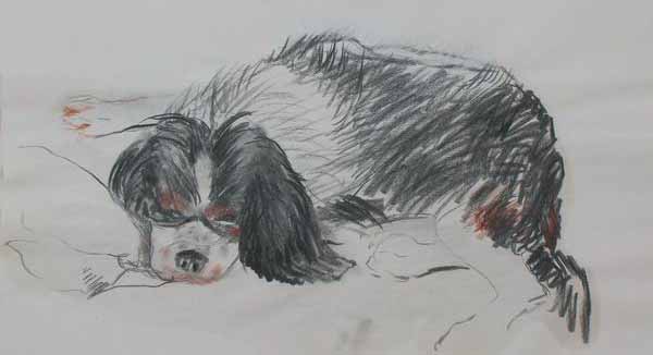 This
article is dedicated to a six year old male cavalier King Charles spaniel named
Callie, who died in June 2006 of heat stroke due to BAOS after a brief walk with
his owner in 79º weather in California, and to a six year old female
cavalier named Bafi (right), who died in September 2007 from BAOS in
Israel.
This
article is dedicated to a six year old male cavalier King Charles spaniel named
Callie, who died in June 2006 of heat stroke due to BAOS after a brief walk with
his owner in 79º weather in California, and to a six year old female
cavalier named Bafi (right), who died in September 2007 from BAOS in
Israel.
RETURN TO TOP
Veterinary Resources
1982-2003
Upper airway obstruction surgery. Harvey C.E. J. Amer. Animal Hosp. Assn. 18: 538–544 (1982). Included CKCS.
Brachycephalic airway obstructive syndrome. Wykes PM. (1991) Problems in Veterinary Medicine 3, 188–197.
"Recognition and treatment of congenital respiratory tract defects in brachycephalics.". Hendricks, JC In Kirk's Current Veterinary Therapy XII Small Animal Practice. p. 892-894. .JD Bonagura and RW Kirk (eds.) 1995. W.B. Saunders Co., Toronto.
Brachycephalic airway obstruction syndrome – a review of 118 cases. Lorinson D., Bright R.M. and White R.A.S. Canine Practice 22: 18–21 (1997). Included CKCS.
Some Practical Solutions to Welfare Problems in Dog Breeding. P D McGreevy and F W Nicholas. Animal Welfare 1999, 8: 329-341. Quote: "As dogs made a transition from working to companion animals, selection for morphological neoteny found favour. This tendency is obvious among old and modem lap dogs. For example, with 'large dark round' eyes, pendant ears and 'compact, cushioned' feet, the Cavalier King Charles Spaniel (Kennel Club, London 1994; FCI Standard No 136) has a very puppy-like conformation."
Temporomandibular joint morphology in Cavalier King Charles spaniels. Alison M. Dickie, Tobias Schwarz, Martin Sullivan. Vet. Rad. & Ultra; May 2002; 43:260
"Brachycephalic airway syndrome." Monnet E. In: Textbook of Small Animal Surgery, 3d Ed. vol. 1, pp.808–813. D Slatter, ed. (2003) WB Saunders, Phila.
RETURN TO TOP
2004-2009
Breed Predispositions to Disease in Dogs & Cats. Alex Gough, Alison Thomas. 2004; Blackwell Publ. 44-45. Quote regarding CKCS: "Brachycephalic upper airway syndrome -- complex of anatomical deformities -- common in this breed -- a likely consequence of selective breeding for certain facial characteristics."
Material in the middle ear of dogs having magnetic resonance imaging for investigation of neurologic signs. Owen MC, Lamb CR, Targett MP. Vet. Radiology & Ultrasound, Mar 2004, 45(2):149-155. Quote: "The aim of this study was to determine the prevalence and potential significance of finding material in the middle ear of dogs having magnetic resonance (MR) imaging. Of 466 MR studies reviewed, an increased signal was identified in the tympanic bulla in 32 (7%) dogs. Cavalier King Charles spaniels, Cocker spaniels, Bulldogs, and Boxers were over-represented compared to the population of dogs having MR imaging. Five (16%) dogs had definite otitis media and one (3%) had a meningioma invading the middle ear. Of the remaining dogs, 13 (41%) had possible otitis media and 13 (41%) had neurologic conditions apparently unrelated to otitis media. The most common appearance of material in the middle ear was isointense in T1-weighted images and hyperintense in T2-weighted images. There was no apparent correlation between the signal characteristics of the material and the diagnosis. Enhanced signal after gadolinium administration was observed affecting the lining of the bulla in dogs with otitis media and in dogs with unrelated neurologic conditions. In dogs without clinical signs of otitis media, finding an increased signal in the middle ear during MR imaging may reflect subclinical otitis media or fluid accumulation unrelated to inflammation. Brachycephalic dogs may be predisposed to this condition."
Brachycephalic Airway Syndrome. Eric Monnet. WSAVA Proceedings 2004
Differences between breeds of dog in a measure of heart rate variability. Doxey S, Boswood A. Vet Rec. 2004 Jun 5;154(23):713-7.
"Brachycephalic airway disease." Dottie Brown, Sue Gregory. In: BSAVA Manual of Canine and Feline Head, Neck and Thoracic Surgery. Daniel Brockman and David Holt, eds. Oct. 2005. Quote: "Breeds particularly predisposed to BAD [brachycephalic airway disease] with elongation of the soft palate include the Bulldog, Pug, Boston Terrier and, in the UK, the Cavalier King Charles Spaniel."
Results of surgical correction of abnormalities associated with brachycephalic airway obstruction syndrome in dogs in Australia. C. V. Torrez and G. B. Hunt. J.Sm.Anim.Prac. March 2006; 47(3):150. Quote: "Stenotic nares were present in 31 dogs (42·5 per cent), elongated soft palate in 63 (86·3 per cent) and everted laryngeal saccules in 43 (58·9 per cent). The most common breeds were the pug (19 dogs, 26 per cent), Cavalier King Charles spaniel (15 dogs, 20·5 per cent), British bulldog (14 dogs, 19·2 per cent) and Staffordshire bull terrier (4 dogs, 5·5 per cent). Laryngeal collapse was present in 34 of 64 (53 per cent) dogs. ... Clinical Significance: Laryngeal collapse is relatively common in dogs presented for surgical correction of brachycephalic airway obstructive disease. Dogs with severe laryngeal collapse often respond well to surgery. Clinical signs rarely resolve completely following surgery."
Surgical correction of brachycephalic syndrome in dogs: 62 cases (1991–2004). Todd W. Riecks, DVM; Stephen J. Birchard, DVM, MS, DACVS; Julie A. Stephens, MS. J.Amer.Vet.Med.Assn. May 1, 2007, 230( 9): 1324-1328. Included CKCS. Quote: "Elongated soft palate was the most common abnormality (54/62 [87.1%] dogs); the most common combination of abnormalities was elongated soft palate, stenotic nares, and everted saccules (16/62 [25.8%] dogs). ... Surgical treatment of brachycephalic syndrome in dogs appeared to be associated with a favorable long-term outcome, regardless of age, breed, specific diagnoses, or number and combinations of diagnoses."
Brachycephalic Syndrome: New Knowledge, New Treatments. Gilles Dupre. 2008 WSAVA Congress. Quote: "Brachycephalic breeds are usually distinguished from others by their shortened skull which results from an early ankylosis of the cartilages in this region. Different breeds are usually recognized as brachycephalic: Boston terrier, English and French bulldog, pugs, Pekinese, Shih-tzu and cavalier King Charles."
RETURN TO TOP
2010
Localization of Canine Brachycephaly Using an Across Breed Mapping Approach. Danika Bannasch, Amy Young, Jeffrey Myers, Katarina Truvé, Peter Dickinson, Jeffrey Gregg, Ryan Davis, Eric Bongcam-Rudloff, Matthew T. Webster, Kerstin Lindblad-Toh, Niels Pedersen. PLOS One. March 2010. Quote: "The domestic dog, Canis familiaris, exhibits profound phenotypic diversity and is an ideal model organism for the genetic dissection of simple and complex traits. However, some of the most interesting phenotypes are fixed in particular breeds and are therefore less tractable to genetic analysis using classical segregation-based mapping approaches. We implemented an across breed mapping approach using a moderately dense SNP array, a low number of animals and breeds carefully selected for the phenotypes of interest to identify genetic variants responsible for breed-defining characteristics. Using a modest number of affected (10–30) and control (20–60) samples from multiple breeds, the correct chromosomal assignment was identified in a proof of concept experiment using three previously defined loci; hyperuricosuria, white spotting and chondrodysplasia. Genome-wide association was performed in a similar manner for one of the most striking morphological traits in dogs: brachycephalic head type. Although candidate gene approaches based on comparable phenotypes in mice and humans have been utilized for this trait, the causative gene has remained elusive using this method. Samples from nine affected breeds and thirteen control breeds identified strong genome-wide associations for brachycephalic head type on Cfa 1. Two independent datasets identified the same genomic region. Levels of relative heterozygosity in the associated region indicate that it has been subjected to a selective sweep, consistent with it being a breed defining morphological characteristic. Genotyping additional dogs in the region confirmed the association. To date, the genetic structure of dog breeds has primarily been exploited for genome wide association for segregating traits. These results demonstrate that non-segregating traits under strong selection are equally tractable to genetic analysis using small sample numbers."
Relationship between pharyngeal conformation and otitis media with effusion in Cavalier King Charles spaniels. Hayes GM, Friend EJ, Jeffery ND. Vet Rec. 2010 Jul 10;167(2):55-8. Quote: "Otitis media with effusion (OME) is a common incidental finding in otherwise normal Cavalier King Charles spaniels (CKCS). ... The incidence of OME as an incidental finding in a sample of 68 CKCS undergoing MRI was 54 per cent in this study, which is comparable to previously reported incidences of 47 per cent ... and 28 per cent ... in this breed. The CKCS in this study were reported to be ‘clinically normal’ by their owners, who did not report clinical signs of neurological disease or otitis in these dogs. ... In this study, measurements made on MRI were used to determine whether there was an association between OME and brachycephalic conformation. The results confirm that association and also demonstrate that, in CKCS, greater thickness of the soft palate and reduced naso-pharyngeal aperture are significantly associated with OME. ... An overlong soft palate has long been accepted as contributing to the [brachycephalic airway] syndrome, but more recently the importance of abnormally thick soft palates has also been recognised ... [B]rachycephalic airway syndrome may occur as a consequence of the selection for morphological neotony in this breed ... Although the aetiology is probably multifactorial, OME is more frequently found in patients with more severe anomalies of nasopharyngeal conformation. Changes within the nasopharynx may impair auditory tube drainage. ... These results suggest that auditory tube dysfunction and OME may represent a previously overlooked consequence of brachycephalic conformation in dogs."
Ocular conditions affecting the brachycephalic breeds. Peter G.C. Bedford. RVC. 2010. Quote: "There are two types of disease which affect the eye of the brachycephalic breeds and both are directly or indirectly related to genetic predisposition. First and by far the commonest are those conditions which are due to be conformation of skull and are related to the exophthalmos which is the common feature of these breeds. Second there are those conditions which have been unwittingly bred into some brachycephalic breeds in the pursuit of desired breed characteristics. In this lecture I will present an overview of all the diseases that the small animal practitioner is likely to encounter in the brachycephalic breeds of pedigree dog. The fourteen breeds I have included for discussion are the Affenpinscher, Boston Terrier, Boxer, Bulldog, Cavalier King Charles and the King Charles Spaniels, (mesaticephalic) French Bulldog, Griffon Bruxellois, Japanese Chin, Lhasa Apso, Pekingese, Pug, Shih Tzu and Tibetan Spaniel. ... Corneal Lipid Dystrophy: The term applies to the characteristic cholesterol and triglyceride deposits in the superficial corneal stroma seen most commonly in the Cavalier King Charles Spaniel. It is clinically benign and seldom affects vision to any noticeable degree. ... Hereditary Cataract: Hereditary cataract is seen in the Boston Terrier and the Cavalier King Charles Spaniel. ... Microphthalmos (MoD): Again the American literature suggests that microphthalmos (MoD) may be inherited in the Cavalier King Charles Spaniel."
Brachycephalic Airway Syndrome Surgery: a Retrospective Analysis of Breeds and Complications in 155 Dogs. Joy-Maree Wetzel, Philip Moses. ANZCVS 2010 Science Week. Quote: "... The most common breeds were the Cavalier King Charles Spaniel (CKCS) (29 dogs, 18.7%) ... All CKCS had an elongated soft palate and accounted for 41% of the laryngeal collapse cases. ..."
Breed Predispositions to Disease in Dogs & Cats (2d Ed.). Alex Gough, Alison Thomas. 2010; Wiley-Blackwell Publ. 54.
Brachycephalic Airway Obstructing Syndrome: Some Further Controversies. Robert N. White. Assn. of Veterinary Soft Tissue Surgeons. Oct. 2010. Quote: "There are a number of breeds in which some individuals show signs consistent with BAOS even though the breeds themselves are not brachycephalic. Examples include the Yorkshire terrier, the Norfolk terrier and the Norwich terrier. Affected individuals rarely show the nasal aspects of the disease recognised in the true brachycephalic breeds. Rather, these breeds present with a range of issues mostly associated with their caudal nasopharynx, pharynx and larynx. ... The Cavalier King Charles spaniel also warrants further discussion. Previous reports from the UK and Australia would suggest that this breed in commonly presented for the investigation and management of BAOS (Lorinson and others 1997, Torrez and Hunt 2006) in both these countries. Surprisingly, a recent retrospective series of 62 dogs from North America (Riecks and others 2007) contained no individuals of the breed. Like the three breeds discussed above [Yorkshire terrier, the Norfolk terrier and the Norwich terrier], it is my experience that the Cavalier King Charles spaniel presented for the further investigation of ʻBAOSʼ commonly does not have the classic findings seen in the bulldog, French bulldog, Pekinese, Pug, etc. Their nasopharyngeal obstruction is often characterised by a subjectively narrow (smaller than expected for a breed of their size) nasopharyngeal space and it is quite common that they do not show evidence of overlength of the soft palate. Interestingly, a recent report (Hayes and others 2010) confirmed a significant association between the presence of otitis media with effusion (on MRI) and an increase thickness of the soft palate and reduced nasopharyngeal aperture. Many of the cases show evidence of otitis media with effusion (OME) on either otoscopic examination or other imaging studies of the tympanic bullae. Malformation of the nasopharynx and soft palate is recognised to be associated with the formation of otitis media with effusion in the dog (White and others 2009). The prevalence of caudal fossa (craniocervical junction abnormalities including occipital hypoplasia) malformations is high in the Cavalier King Charles spaniel and is considered to be associated with the presence of neurological signs observed in individuals suffering from cerebellar herniation and syringohydromyelia (Cerda-Gonzalez and others 2009). It would seem reasonable to hypothesise that the Cavalier King Charles spaniel might suffer from a syndrome of conditions (syringomyelia, OME, nasopharyngeal airway obstruction, etc.) that are all associated with the malformation of the caudal aspect of the skull."
Brachycephalic airway obstructive syndrome in dogs: 90 cases (1991–2008). Frank J. Fasanella, Jacob M. Shivley, Jennifer L. Wardlaw, Sumalee Givaruangsawat. JAVMA Nov 2010; 237(9): 1048-1051. Quote: "Objective—To determine the prevalence of individual anatomic components of brachycephalic airway obstructive syndrome (BAOS), including everted tonsils, and analyze the frequency with which each component occurs with 1 or more other components of BAOS in brachycephalic dogs. ... 90 dogs with BAOS. Results—English Bulldogs (55/90 [61%]), Pugs (19/90 [21%]), and Boston Terriers (8/90 [9%]) were the most common breeds with BAOS. The most common components of BAOS were elongated soft palate (85/90 [94%]), stenotic nares (69/90 [77%]), everted laryngeal saccules (59/90 [66%]), and everted tonsils (50/90 [56%]). Dogs most commonly had 3 or 4 components of BAOS, with the most common combination being stenotic nares, elongated soft palate, everted laryngeal saccules, and everted tonsils. Dogs with stenotic nares were significantly more likely to have everted laryngeal saccules (50/69 [72%]), and dogs with everted laryngeal saccules were significantly more likely to have everted tonsils (39/59 [66%]). Postoperative surgical complications occurred in 12% (10/83) of dogs that received corrective surgery. No specific BAOS component made dogs more likely to have complications. Conclusions and Clinical Relevance—The prevalence of components of BAOS in brachycephalic dogs of this study differed from that reported previously, especially for everted tonsils. Thorough examination of the pharynx and larynx is necessary for detection of BAOS components."
Human evolutionary history: Consequences for the pathogenesis of otitis media. Charles D Bluestone, J. Douglas Swarts. Otolaryngology -- Head & Neck Surgery; Dec 2010; 143(6):739-744. Quote: "Among veterinarians, chronic ME effusion (termed primary secretory otitis media) is a well-known disease in the Cavalier King Charles Spaniel. It has been reported to be present in up to 40 percent of these animals. The effusion is mucoid and fills the entire ME. Diagnosis is made by operating microscopic examination, computed tomography scanning, or magnetic resonance imaging (MRI), and has been confirmed at the time of myringotomy. Myringotomy and tympanostomy tube placement has been recommended for treatment. This breed has been artificially selected to have a shortened front-to-back diameter of the skull, a shape termed brachycephaly, which arises due to premature fusion of the coronal sutures. The term neotenous (retention of juvenile characteristics into adulthood) is also appropriate for these breeds. The Cavalier snores habitually like other brachycephalic dogs, including the English Bulldog, a breed that has been reported to be the only animal known to develop obstructive sleep apnea. The snoring is undoubtedly secondary to its constricted pharynx, a consequence of the shortening of the snout. ... Figure 5 compares the head shape of a Cavalier King Charles Spaniel, with its extremely short face, to that of a Golden Retriever, which has a classic prognathic snout. The Cavalier King Charles Spaniel is an animal model of chronic OM with effusion. It has been “artificially selected” (Charles Darwin's term) for its short snout and globular head, but an unintended consequence of breeding for this characteristic is the propensity for chronic OM with effusion. In a recently reported study using MRI, veterinarians from England found that not only did the Cavalier have OM (54%), but another brachycephalic breed, the Boxer, also had ME disease (32%), which was not present in Cocker Spaniels, a mesaticephalic breed. The investigators suggested that the reduced nasopharyngeal space in the Cavalier and Boxers, when compared with the Cocker Spaniel, predisposed them to OM. It might be that one or both of the paratubal muscles is dysfunctional due to the abnormal palatal anatomy in these breeds and is the cause of their OM. The underlying pathogenesis of the Cavalier's ME disease is currently under investigation in our laboratory. Analogously, one could speculate that with the loss of their prognathic face, humans became susceptible to OM, an unintended consequence of “natural selection” for another adaptation (again, Darwin's term), as described above."
RETURN TO TOP
2011
Cephalometric Measurements and Determination of General Skull Type of Cavalier King Charles Spaniels. M. J. Schmidt, A. C. Neumann, K. H. Amort, K. Failing, M. Kramer. Vet. Rad. & Ultra, 26 Apr 2011. Quote: "The general skull morphology of the head of the Cavalier King Charles Spaniel (CKCS) was examined and compared with cephalometric indices of brachycephalic, mesaticephalic, and dolichocephalic heads. Measurements were taken from computed tomography images. Defined landmarks for linear measurements of were identified using three-dimensional (3D) models. The calculated parameters of the CKCS were different from all parameters of mesaticephalic dogs but were the same as parameters from brachycephalic dogs. However, the CKCS had a wider braincase in relation to length than in other brachycephalic breeds. Studies of the etiology of the chiari-like malformation in the CKCS should therefore focus on brachycephalic control groups. As Chari-like malformation has only been reported in brachycephalic breeds, its etiology could be associated with a higher grade of brachycephaly, meaning a shorter longitudinal extension of the skull. This has been suggested for other breeds."
Canine brachycephalic airway syndrome: pathophysiology, diagnosis, and nonsurgical management. Michelle Trappler, Kenneth W. Moore. Compend Contin Educ Vet. May 2011;33(5):E1-4; quiz E5. Quote: Canine brachycephalic airway syndrome is a progressive disease that affects many brachycephalic dogs. This article describes the components of this syndrome and focuses on acute emergency management and long-term conservative management of these patients. Surgical management is described in a companion article.
Bronchomalacia in Dogs with Myxomatous Mitral Valve Degeneration. MK Singh, LR Johnson, MD Kittleson, RE Pollard. William R. Pritchard. J Vet Intern Med 2011;--- (ACVIM 29th Ann. Vet. Med. Forum Abstract Program: Abstract C-12). Quote: "Coughing in the small breed dog may be related to cardiac causes associated with myxomatous mitral valve degeneration (MMVD) including pulmonary edema and compression of the mainstem bronchus by a severely enlarged left atrium, or due to respiratory causes such as tracheal and/or bronchial collapse or chronic bronchitis. The purpose of this study was to evaluate the association between left atrial enlargement and large airway collapse in dogs with MMVD and chronic cough. We hypothesized that airway collapse was independent of degree of left atrial enlargement. ... Preliminary results failed to identify an association between left atrial enlargement and airway collapse in dogs with MMVD but did suggest that airway inflammation is common in affected dogs. Further studies are needed to identify factors contributing to airway collapse in dogs with and without MMVD."
Canine Inherited Disorders Database: http://ic.upei.ca/cidd/disorder/brachycephalic-syndrome
The Anatomy of the Dog Soft Palate. II. Histological Evaluation of the Caudal Soft Palate in Brachycephalic Breeds With Grade I Brachycephalic Airway Obstructive Syndrome. Michela Pichetto, Silvana Arrighi, Paola Roccabianca, Stefano Romussi. Anatomical Record. July 2011;294(7):1267-1272. Quote: "In brachycephalic dogs, the skull bone shortening is not paralleled by a decreased development of soft tissues. Relatively longer soft palate is one of the main factors contributing to pharyngeal narrowing during normal respiratory activity of these dog breeds, which are frequent carriers of the brachycephalic airway obstructive syndrome (BAOS), which affects most part of them during their postnatal life. No histological studies assessing the morphology and the normal tissue composition of the soft palate in brachycephalic dogs are available, neither has ever been determined whether the elongated soft palate is a primary or secondary event. Aim of this study was to describe the morphology of the caudal soft palate in brachycephalic dogs with Grade I BAOS to identify potential features possibly favoring the pathogenesis of BAOS. Specimens from brachycephalic dogs (N = 11) that underwent preventive surgery were collected from surgery, processed for histology, and examined at six transversal levels. The brachycephalic soft palates showed peculiar features such as thickened superficial epithelium, extensive oedema of the connective tissue, and mucous gland hyperplasia. Several muscular alterations were evidenced in addition. The results of this investigation add to the general knowledge of the anatomy of soft palate in the canine species and establish baseline information on the morphological basis of the soft palate thickening in brachycephalic dogs."
Underlying diseases in dogs referred to a veterinary teaching hospital because of dyspnoea: 229 cases (2003-2007). Sonja Fonfara, Lourdes de la Heras Alegret, Alexander J. German, Laura Blackwood, Joanna Dukes-McEwan, P-J. M. Noble, Rachel D. Burrow. J.Am.Vet.Med.Assn.; Nov 2011; 239(9):1219-1224. Quote: "Objective—To identify the most frequent underlying diseases in dogs examined because of dyspnea and determine whether signalment, clinical signs, and duration of clinical signs might help guide assessment of the underlying condition and prognosis. Design—Retrospective case series. Animals—229 dogs with dyspnea. (Bulldogs, Cavalier King Charles Spaniels, Staffordshire Bull Terriers, Yorkshire Terriers and Pugs were overrepresented in the dyspnoeic population.) Results—Upper airway (n = 74 [32%]) and lower respiratory tract (76 [33%]) disease were the most common diagnoses, followed by pleural space (44 [19%]) and cardiac (27 [12%]) diseases. Dogs with upper airway and pleural space disease were significantly younger than dogs with lower respiratory tract and cardiac diseases. Dogs with lower respiratory tract and associated systemic diseases were significantly less likely to be discharged from the hospital. Dogs with diseases that were treated surgically had a significantly better outcome than did medically treated patients, which were significantly more likely to be examined on an emergency basis with short duration of clinical signs. Conclusions and Clinical Relevance—In dogs examined because of dyspnea, young dogs may be examined more frequently with breed-associated upper respiratory tract obstruction or pleural space disease after trauma, whereas older dogs may be seen more commonly with progressive lower respiratory tract or acquired cardiac diseases. Nontraumatic acute onset dyspnea is often associated with a poor prognosis, but stabilization, especially in patients with cardiac disease, is possible. Obesity can be an important contributing or exacerbating factor in dyspneic dogs."
Use of the harmonic scalpel for soft palate resection in dogs: a series of three cases. J Michelsen. Austr Vet J; Nov 2011; 89(12):511-514. Quote: Case 2: A 7-year-old male neuter Cavalier King Charles Spaniel was presented with moderate respiratory compromise secondary to BAOS. The patient had a history of slowly worsening inspiratory obstruction and was becoming increasingly exercise intolerant. The nares were not stenotic and the laryngeal saccules appeared normal. Under general anaesthesia it was judged that the soft palate was 5 mm too long and surgery was performed to resect the redundant tissue. The staphylectomy took 5 min without complications. The modified surgical procedure was used in this case and the patient recovered uneventfully, with no postoperative bleeding or respiratory compromise. Respiratory function was much improved at recovery from anaesthesia and at 24 h. The patient was virtually asymptomatic 6 months postoperatively. ... Soft palate resection is performed to resect a redundant or diseased soft palate, often associated with brachycephalic airway obstructive syndrome (BAOS). Resection has been associated with numerous complications, including coughing, bleeding, pharyngeal oedema, respiratory obstruction and death. Traditionally, the surgery is performed by sharp dissection and suturing, but other reported techniques include the use of an electrothermal sealing device or a laser. Operative time for sharp dissection is approximately 12 min, but is shortened to around 5 min when using a laser, as the haemostatic properties of the instrument negates the need for post-resection oversewing. The successful use of a harmonic scalpel to resect redundant soft palates in three dogs is described. The resected soft palates were not oversewn and the surgical time was comparable with that for laser surgery. The first dog had a minor bleed 6 h postoperatively, possibly associated with suboptimal placement of the harmonic scalpel cutting jaws. The following two patients had no postoperative complications. The harmonic scalpel laparoscopic handpiece allowed excellent visualisation of the surgical field and rapid performance of the procedure. All three patients had markedly improved postoperative respiratory function. Cleaning and resterilisation permitted multiple reuse of the handpiece, making it cost-competitive with other surgical techniques.
RETURN TO TOP
2012
Surgical management of laryngeal collapse associated with brachycephalic airway obstruction syndrome in dogs. R. N. White. J Sm An Prac; Jan 2012;53(1):44–50. Quote: "Objective: To describe the use of cricoarytenoid lateralisation combined with thyroarytenoid caudo-lateralisation (arytenoid laryngoplasty) for the management of stage II and III laryngeal collapse in dogs. Methods: A retrospective study of a consecutive series of 12 dogs [five breeds were represented; Cavalier King Charles spaniel (n=3), English bulldog (n=4), French bull-dog (n=2), pug (n=2) and Pekingese] suffering from life-threatening stage II or III laryngeal collapse associated with brachycephalic airway obstruction syndrome. Results: Pre-operatively, either stage II collapse (2/12) or stage III collapse (10/12) was confirmed on visual examination. In all cases, a left-sided arytenoid laryngoplasty was performed. Two dogs were euthanased postoperatively as a result of persistent life-threatening respiratory compromise. The procedure resulted in subjective enlargement of the rima glottidis and an associated improvement in respiratory function in the remaining 10 dogs. Follow-up, long-term outcome (median, 3·5 years) in these dogs indicated that all owners considered that the surgery had resulted in marked improvements in their dog's respiratory function, tolerance to exercise, and quality of life. Clinical Significance: Combined cricoarytenoid and thyroarytenoid caudo-lateralisation may be a useful procedure for treatment of stage II and III laryngeal collapse in the dog."
Breed-Specific Anesthesia. Stephanie Krein, Lois A. Wetmore. NAVA Clinicians Brief; March 2012; 17-20. Quote: "Certain breed differences can lead to greater risks for airway obstruction, increased responsiveness to anesthetic drugs, and delayed recovery, all of which can result in increased anesthesia-related morbidity and mortality. ... If cardiac disease is suspected, a full cardiac workup with a veterinary cardiologist is recommended. ... Brachycephalic Breeds (e.g. bulldog, pug, Boston terrier, boxer, Cavalier King Charles spaniel, Pekingese). Problem: Brachycephalic airway syndrome; increased respiratory effort; potential for upper airway obstruction. Avoid excessive sedation. Avoid α2-agonists. Administer acepromazine at half dose. Preoxygenate. Use short-acting induction agent. Use appropriately sized endotracheal tubes. Extubate after patient is sitting up, vigorously chewing, bright, alert. ... Brachycephalic breeds have anatomic considerations that may affect anesthetic outcome.Most brachycephalic breeds suffer from brachycephalic airway syndrome (BAS), which is characterized by stenotic nares, elongated soft palate, everted laryngeal saccules, and hypoplastic trachea. Affected dogs have narrower upper airways than do dogs with normal anatomic features. Because in brachycephalic breeds additional airway contraction can occur with stress (ie, increased respiratory effort, turbulent flow), clinicians need to be prepared for possible upper airway obstruction. Furthermore, brachycephalic dogs must be monitored closely after premedication, throughout anesthesia and the postoperative period, and after extubation. An oxygen source and endotracheal tube should be readily available. Many brachycephalic dogs respond well to acepromazine in conjunction with an opioid; however, the sedative dose should be half of that used for nonbrachycephalic dogs. Full mu-opioid agonists can be used but because they may cause excessive respiratory depression, a reversal agent should be available. Dexmedetomidine should be avoided because of the presence of high vagal tone in these breeds. Anticholinergics, such as glycopyrrolate, may be used to decrease airway secretions and counteract high vagal tone. Preoxygenation is recommended before dogs with BAS are induced. Propofol or a similar short-acting drug should be used for induction and intubation should be completed as rapidly as possible.Mask inductions should be avoided, and smaller endotracheal tubes should be used. Because brachycephalic breeds tend toward obesity, controlled or mechanical ventilation is often necessary.Most problems associated with mechanical ventilation occur during induction and recovery, so monitoring is particularly important. Extubation should be postponed until the patient is bright, alert, swallowing—even chewing on the endotracheal tube. If extubation is attempted while the patient is sedated and groggy from anesthesia, there is increased risk for upper airway obstruction. If upper airway obstruction occurs, the patient should be reintubated."
Do dog owners perceive the clinical signs related to conformational inherited disorders as ‘normal’ for the breed? A potential constraint to improving canine welfare. RMA Packer A Hendricks, and CC Burn. Animal Welfare May 2012; 21(S1): 81-93. Quote: "Selection for brachycephalic (foreshortened muzzle) phenotypes in dogs is a major risk factor for brachycephalic obstructive airway syndrome (BOAS). Clinical signs include respiratory distress, exercise intolerance, upper respiratory noise and collapse. Efforts to combat BOAS may be constrained by a perception that it is ‘normal’ in brachycephalic dogs. This study aimed to quantify owner perception of the clinical signs of BOAS as a veterinary problem. A questionnaire-based study was carried out over five months on the owners of dogs referred to the Queen Mother Hospital for Animals (QMHA) for all clinical services, except for Emergency and Critical Care. Owners reported the frequency of respiratory difficulty and characteristics of respiratory noise in their dogs in four scenarios, summarised as an ‘owner-reported breathing’ (ORB) score. Owners then reported whether their dog currently has, or has a history of, ‘breathing problems’. Dogs (n = 285) representing 68 breeds were included, 31 of which were classed as ‘affected’ by BOAS either following diagnostics, or by fitting case criteria based on their ORB score, skull morphology and presence of stenotic nares [including cavalier King Charles spaniels]. The median ORB score given by affected dogs’ owners was 20/40 (range 8–30). Over half (58%) of owners of affected dogs reported that their dog did not have a breathing problem. This marked disparity between owners’ reports of frequent, severe clinical signs and their perceived lack of a ‘breathing problem’ in their dogs is of concern. Without appreciation of the welfare implications of BOAS, affected but undiagnosed dogs may be negatively affected indefinitely through lack of treatment. Furthermore, affected dogs may continue to be selected in breeding programmes, perpetuating this disorder."
Normal for the breed? Rowena Packer. Vet. Nurse. June 2012;3(5):326. Quote: "Brachycephalic dogs are at high-risk of brachycephalic obstructive airway syndrome (BOAS). Clinical signs include noisy and laboured breathing, breathing difficulties even on short walks and easily overheating. These difficulties can prevent dogs from being able to enjoy simple pleasures such as exercise, play, food and sleep. In severe cases dogs can experience almost continuous breathing difficulties and collapse due to lack of oxygen. Due to the chronic and prevalent nature of clinical signs, they may be ‘accepted’ by owners and not perceived as abnormal, with only particularly acute or severe attacks alerting owners to present their dog to their veterinary practice. This is of additional concern as clinical signs often get worse over time if they are left untreated, and prognosis is improved with early intervention. ... Without appreciation of the welfare implications of BOAS, affected but undiagnosed dogs may be negatively affected indefinitely through lack of treatment. As such, this is an area that the veterinary profession should aim to tackle by raising awareness through client education. For example, we urge the veterinary team to make clients aware of what clinical signs such as snoring, snorting and difficulty exercising may indicate in these breeds, even if their dogs are not presented for these signs but are suspected to have BOAS."
Effect of brachycephalic, mesaticephalic, and dolichocephalic head conformations on olfactory bulb angle and orientation in dogs as determined by use of in vivo magnetic resonance imaging. Aseel K. Hussein, Martin Sullivan, Jacques Penderis. Am.J.Vety.Res. July 2012; 73(7):946-951. Quote: "Objective: To determine the effect of head conformation (brachycephalic, mesaticephalic, and dolichocephalic) on olfactory bulb angle and orientation in dogs by use of in vivo MRI. Animals: 40 client-owned dogs undergoing MRI for diagnosis of conditions that did not affect skull conformation or olfactory bulb anatomy. Procedures: For each dog, 2 head conformation indices were calculated. Olfactory bulb angle and an index of olfactory bulb orientation relative to the rest of the CNS were determined by use of measurements obtained from sagittal T2-weighted MRI images. Results: A significant negative correlation was found between olfactory bulb angle and values of both head conformation indices. Ventral orientation of olfactory bulbs was significantly correlated with high head conformation index values (ie, brachycephalic head conformation). Conclusions and Clinical Relevance: Low olfactory bulb angles and ventral olfactory bulb orientations were associated with brachycephalia. Positioning of the olfactory bulbs, cribriform plate, and ethmoid turbinates was related. Indices of olfactory bulb angle and orientation may be useful for identification of dogs with extremely brachycephalic head conformations. Such information may be used by breeders to reduce the incidence or severity of brachycephalic-associated diseases."
Brachycephalic Airway Syndrome: Pathophysiology and Diagnosis. Dena L. Lodato, Cheryl S. Hedlund. Compendium. July 2012; 34(7).
Brachycephalic Airway Syndrome: Management. Dena L. Lodato, Cheryl S. Hedlund. Compendium. Aug 2012; 34(8).
Variation of BMP3 Contributes to Dog Breed Skull Diversity. Jeffrey J. Schoenebeck, Sarah A. Hutchinson, Alexandra Byers, Holly C. Beale, Blake Carrington, Daniel L. Faden, Maud Rimbault, Brennan Decker, Jeffrey M. Kidd, Raman Sood, Adam R. Boyko, John W. Fondon, III, Robert K. Wayne, Carlos D. Bustamante, Brian Ciruna, Elaine A. Ostrander. PLoS Genet. 2012 August; 8(8): e1002849. Quote: "As a result of selective breeding practices, modern dogs display a multitude of head shapes. Breeds such as the Pug and Bulldog popularize one of these morphologies, termed “brachycephaly.” A short, upward-pointing snout, a massive and rounded head, and an underbite typify brachycephalic breeds. Here, we have coupled the phenotypes collected from museum skulls with the genotypes collected from dogs and identified five regions of the dog genome that are associated with canine brachycephaly [including the cavalier King Charles spaniel]. Fine mapping at one of these regions revealed a causal mutation in the gene BMP3. Bmp3's role in regulating cranial development is evolutionarily ancient, as zebrafish require its function to generate a normal craniofacial morphology. Our data begin to expose the genetic mechanisms unknowingly employed by breeders to create and diversify the cranial shape of dogs."
Do dog owners perceive the clinical signs related to conformational inherited disorders as ‘normal’ for the breed? A potential constraint to improving canine welfare. (Owner recognition of breathing disorders in brachycephalic dogs). RMA Packer, A Hendricks and CC Burn. Animal Welfare. 2012;21(S1):81-93. Quote: "Selection for brachycephalic (foreshortened muzzle) phenotypes in dogs is a major risk factor for brachycephalic obstructive airway syndrome (BOAS). Clinical signs include respiratory distress, exercise intolerance, upper respiratory noise and collapse. Efforts to combat BOAS may be constrained by a perception that it is ‘normal’ in brachycephalic dogs. This study aimed to quantify ownerperception of the clinical signs of BOAS as a veterinary problem. A questionnaire-based study was carried out over five months on the owners of dogs referred to the Queen Mother Hospital for Animals (QMHA) for all clinical services, except for Emergency and Critical Care. Owners reported the frequency of respiratory difficulty and characteristics of respiratory noise in their dogs in four scenarios, summarised as an ‘owner-reported breathing’ (ORB) score. Owners then reported whether their dog currently has, or has a history of, ‘breathing problems’. Dogs (n = 285) representing 68 breeds were included, 31 of which were classed as ‘affected’ by BOAS either following diagnostics, or by fitting case criteria based on their ORB score, skull morphology and presence of stenotic nares. The median ORB score given by affected dogs’ owners was 20/40 (range 8–30). Over half (58%) of owners of affected dogs reported that their dog did not have a breathing problem. This marked disparity between owners’ reports of frequent, severe clinical signs and their perceived lack of a ‘breathing problem’ in their dogs is of concern. Without appreciation of the welfare implications of BOAS, affected but undiagnosed dogs may be negatively affected indefinitely through lack of treatment. Furthermore, affected dogs may continue to be selected in breeding programmes, perpetuating this disorder."
RETURN TO TOP
2013
Stop brachycephalism, now! Fraser Hale. Can.Vet.J. Feb. 2013;54(2):185-186. Quote: "In many Canadian jurisdictions, veterinarians have advocated for and achieved a ban on tail-docking, ear-cropping, and dewclaw removal as these are considered unnecessary cosmetic procedures that cause (temporary) pain with no benefit to the animals. I believe that as protectors of animal welfare, veterinarians should start a public awareness campaign to inform people of the serious, life-long negative impacts of brachycephalism. I believe we must stop referring to these conditions as 'normal for the breed' and refer to them as 'grossly abnormal in accordance with breed standards' because there is nothing re2013but comical breeds in advertising and promotional materials as this just increases public demand because they are 'so cute.'"
RETURN TO TOP
2014
Laryngeal Disease in Dogs and Cats. Catriona MacPhail. Vet. Clinics of N.A.: Sm. Anim. Prac. Jan. 2014;44(1):19-31. Quote: "Brachycephalic airway syndrome refers to the condition of obstructive airway distress attributable to anatomic abnormalities of breeds such as English and French bulldogs, pugs, Boston terriers, and Cavalier King Charles spaniels."
Assoziation zephalometrischer Parameter mit dem Auftreten der Syringomyelie beim Cavalier King Charles Spaniel mit Chiari-ähnlicher Malformation [Association of anatomical parameters with the occurrence of syringomyelia in the Cavalier King Charles Spaniel with Chiari malformation]. Annabell Johanna Grübmeyer. Giessener Elektronische Bibliothek. Jan. 2014. Quote: "Until now the Chiari-like malformation was only diagnosed in brachycephalic dog breeds. Based on the decreased length-breadth ratio of its skull the Cavalier King Charles Spaniel can be classified as a highly brachycephalic dog. Therefore it could be assumed that the grade of brachycephaly is a pathophysiological factor for the development of syringomyelia and a retarded length growth of the skull might be the cause for the changes found in the Chiari-like malformation. The question is, if a shortening of the cranial base gives rise to the pathological changes in Chiari-like malformation. Based on this question we examined the anatomical parameters of 107 Cavalier King Charles Spaniels in relationship to the occurrence of syringomyelia. The study should give information about the pathogenesis of the Chiari-like malformation and the development of syringomyelia and if there is a difference in the cranial base length in Cavalier King Charles Spaniels with or without syringomyelia. The 107 Cavalier King Charles Spaniels examined in this study were mostly presented for breeding examinations, but some were also presented because of clinical signs. The age of the examined dogs ranged from 6 month to 9 years. We performed computed tomography of the skull and magnetic resonance imaging of the skull and spine of all patients. The examination of the spine in patients introduced for breeding examinations, were performed until the 5th cervical vertebra. In patients with neurological signs the examination included also the caudal cervical, thoracic and lumbar spine. Changes consistent with the Chiari-like malformation were found in all 107 Cavalier King Charles Spaniels. 63 of the 107 dogs showed a syringomyelia at the point of examination. The results of the study showed that the incidence of syringomyelia is correlated to the variables age (p less than 0,007), SBI [skull base index] (p less than 0,0192), PI [presphenoid index] (p less than 0,0447) and BI [basisphenoid index] (p less than 0,0206). Furthermore it is shown that Cavalier King Charles Spaniels with a decrease in SBI have an increased risk to develop syringomyelia (odds ratio 1,26). In addition also the presphenoid and the basioccipital bone showed a reduced length, with an increase in breadth in dogs with syringomyelia. This study showed, that a reduced length of the cranial base represents a risk factor for the occurrence of syringomyelia. These results support the assumption of other authors that the cause of the Chiari-like malformation and syringomyelia is up to a growth disturbance of the cranial base."
Tracheal Collapse in Dogs. Mary Dell Deweese, Karen M. Tobias. Clinicians Brief. May 2014;83-87.
Evaluation of a novel tracheal stent for the treatment of tracheal collapse in dogs. D. Clarke, E. de Madron, R. Presley. J.Vet.Int.Med. July 2014;28(4):1364. Quote: "The purpose of this study was to evaluate the safety, efficacy, and satisfaction associated with a novel variable diameter tracheal stent designed specifically for canine anatomy and tracheal collapse. This was a multicenter retrospective study of 27 consecutive cases of tracheal collapse treated with the stent. Data forms requiring chart review and client interviews were distributed to the veterinarian placing each stent. Information was collected that compared symptoms pre- and post-stenting as well as a number of other parameters relating to the procedure and subsequent clinical course to assess stent performance and ease of use. Symptoms were graded on a visual line scale of 0-10. Paired t-tests or sign rank tests were used to determine changes in symptoms following stenting. A two-sided p-value of <0.05 was considered statistically significant. Responses were obtained from 20 of 27 cases. Data was normally distributed. The mean duration of follow-up was 3 months (range >1 to 17 months). There were no procedural complications. Operative mortality was 0%. There were no stent fractures or stent related deaths; symptomatic granulation tissue developed in one case. Statistically significant improvement was seen in cough, respiratory function, quality of life, and exercise tolerance. All veterinarians stated they would use the stent again. 95% of clients would repeat the procedure and 90% expressed satisfaction with the outcome. Tracheal stenting performed with the novel implant is both safe and subjectively effective in significantly improving clinical symptoms. Veterinarians and clients expressed a positive perception regarding outcomes associated with the stent."
Introducing breathlessness as a significant animal welfare issue. Beausoleil N, Mellor D. N.Z.Vet.J. July 2014;8:1-22. Quote: "Breathlessness is a negative affective experience relating to respiration, the animal welfare significance of which has largely been underestimated in the veterinary and animal welfare sciences. In this review, we draw attention to the negative impact that breathlessness can have on the welfare of individual animals and to the wide range of situations in which mammals may experience breathlessness. At least three qualitatively distinct sensations of breathlessness are recognised in human medicine - respiratory effort, air hunger and chest tightness - and each of these reflects comparison by cerebral cortical processing of some combination of heightened ventilatory drive and/or impaired respiratory function. Each one occurs in a variety of pathological conditions and other situations, and more than one may be experienced simultaneously or in succession. However, the three qualities vary in terms of their unpleasantness, with air hunger reported to be the most unpleasant. We emphasise the important interplay among various primary stimuli to breathlessness and other physiological and pathophysiological conditions, as well as animal management practices. For example, asphyxia/drowning of healthy mammals or killing those with respiratory disease using gases containing high carbon dioxide tensions is likely to lead to severe air hunger, while brachycephalic obstructive airway syndrome in modern dog and cat breeds increases respiratory effort at rest and likely leads to air hunger during exertion. Using this information as a guide, we encourage animal welfare scientists, veterinarians, laboratory scientists, regulatory bodies and others involved in evaluations of animal welfare to consider whether or not breathlessness contributes to any compromise they may observe or wish to avoid or mitigate."
Brachycephalic obstructive airway syndrome: a growing problem. Terry Emmerson. J. Small Animal Practice. November 2014;55(11):543-544. Quote: "The Cavalier King Charles spaniel (CKCS) is often considered a brachycephalic breed and can present with typical brachycephalic airway signs such as respiratory noise, snoring, stertor and exercise intolerance. They are represented in reviews of dogs undergoing corrective airway surgery for BOAS and dogs requiring tracheostomies (Torrez & Hunt 2006). However these dogs often do not demonstrate the typical abnormalities of the brachycephalic breeds. They often do not have an overlong palate as judged by standard criteria or stenotic nares or everted laryngeal ventricles. Some present with isolated laryngeal collapse and we have seen some dogs with concurrent laryngeal paralysis. As a breed they often have a particularly thick soft palate and small nasopharynx which may be the main factors driving the signs in this breed. Other breeds that can present with BOAS-like symptoms but often without the typical abnormalities include the bull terrier and Mastiff breeds. Like the CKCS, these breeds often do not have a surgically correctable problem. BOAS is a complex problem which currently we do not fully understand. We need to better understand the anatomical and breed variations in these cases and their relationship to severity of signs and postsurgical outcome. Current surgical options offer a way to alleviate signs and can provide significant improvement. However, there is still a frustrating subset of cases with poor outcomes that we struggle to identify pre-operatively. Surgery should be performed on affected dogs to improve quality of life and as a welfare issue. However, ultimately, the only way to truly improve this condition is by education of the public and breeders regarding the problems of these breeds and with breeding aimed at improving conformation."
Use of silicone tracheal stoma stents for temporary tracheostomy in dogs with upper airway obstruction. T. Trinterud, P. Nelissen, R. A. S. White. J. Small Animal Practice. November 2014;55(11):551-559. Quote: "Objectives: To report the use of silicone tracheal stoma stents for temporary tracheostomy in dogs with upper airway obstruction. Methods: Retrospective review of medical records for dogs in which silicone tracheal stoma stents were placed. Results: Eighteen dogs [including three cavalier King Charles spaniels] had a silicone tracheal stoma stent placed for maintenance of a tracheostomy stoma for periods ranging from three hours to eight months. No intra-operative or immediate postoperative complications were recorded. In 11 dogs the stent was removed by simple traction after a period ranging from 36 hours to 6 weeks, and the tracheal stoma was left to heal by second intention. Five of the 18 dogs were determined as being tracheostomy dependent and underwent conversion to permanent tracheostomy after a period ranging from five days to eight months following stent placement. One dog was euthanased after three months, with the stent still in place, because of poor respiratory function, and one dog [a CKCS] died of unrelated reasons ["severe immune-mediated thrombocytopenia (ITP)"]. In 6 of 10 dogs (60%) where the stent was in place for five days or more, granulation tissue formation caused dislodgement of the stent. Clinical Significance: Silicone tracheal stoma stents may be used as an alternative to conventional trache-ostomy tubes in selected dogs with upper airway obstruction. Long-term use of the stent beyond five days is not recommended because of granulation tissue formation. The long-term consequences of partial tracheal ring resection are unknown."
RETURN TO TOP
2015
Introducing breathlessness as a significant animal welfare issue. NJ Beausoleila, DJ Mellora. N. Z. Vet. J. January 2015;63(1):44-51. Quote: Breathlessness is a negative affective experience relating to respiration, the animal welfare significance of which has largely been underestimated in the veterinary and animal welfare sciences. In this review, we draw attention to the negative impact that breathlessness can have on the welfare of individual animals and to the wide range of situations in which mammals may experience breathlessness. At least three qualitatively distinct sensations of breathlessness are recognised in human medicine--respiratory effort, air hunger and chest tightness--and each of these reflects comparison by cerebral cortical processing of some combination of heightened ventilatory drive and/or impaired respiratory function. Each one occurs in a variety of pathological conditions and other situations, and more than one may be experienced simultaneously or in succession. However, the three qualities vary in terms of their unpleasantness, with air hunger reported to be the most unpleasant. We emphasise the important interplay among various primary stimuli to breathlessness and other physiological and pathophysiological conditions, as well as animal management practices. For example, asphyxia/drowning of healthy mammals or killing those with respiratory disease using gases containing high carbon dioxide tensions is likely to lead to severe air hunger, while brachycephalic obstructive airway syndrome in modern dog and cat breeds increases respiratory effort at rest and likely leads to air hunger during exertion. Using this information as a guide, we encourage animal welfare scientists, veterinarians, laboratory scientists, regulatory bodies and others involved in evaluations of animal welfare to consider whether or not breathlessness contributes to any compromise they may observe or wish to avoid or mitigate.
Comparison of the Relationship between Cerebral White Matter and Grey Matter in Normal Dogs and Dogs with Lateral Ventricular Enlargement. Martin J. Schmidt, Steffi Laubner, Malgorzata Kolecka, Klaus Failing, Andreas Moritz, Martin Kramer, Nele Ondreka. PLoS ONE 10(5): e0124174. May 2015. Quote: "Large cerebral ventricles are a frequent finding in brains of dogs with brachycephalic skull conformation, in comparison with mesaticephalic dogs. It remains unclear whether oversized ventricles represent a normal variant or a pathological condition in brachycephalic dogs. There is a distinct relationship between white matter and grey matter in the cerebrum of all eutherian mammals. The aim of this study was to determine if this physiological proportion between white matter and grey matter of the forebrain still exists in brachycephalic dogs with oversized ventricles. The relative cerebral grey matter, white matter and cerebrospinal fluid volume in dogs were determined based on magnetic-resonance-imaging datasets using graphical software. In an analysis of covariance (ANCOVA) using body mass as the covariate, the adjusted means of the brain tissue volumes of two groups of dogs were compared. Group 1 included 37 mesaticephalic dogs of different sizes with no apparent changes in brain morphology, and subjectively normal ventricle size. Group 2 included 35 brachycephalic dogs [including 7 cavalier King Charles spaniels, 4 of which were diagnosed with syringomyelia] in which subjectively enlarged cerebral ventricles were noted as an incidental finding in their magnetic-resonance-imaging examination. Whereas no significant different adjusted means of the grey matter could be determined, the group of brachycephalic dogs had significantly larger adjusted means of lateral cerebral ventricles and significantly less adjusted means of relative white matter volume. This indicates that brachycephalic dogs with subjective ventriculomegaly have less white matter, as expected based on their body weight and cerebral volume. Our study suggests that ventriculomegaly in brachycephalic dogs is not a normal variant of ventricular volume. Based on the changes in the relative proportion of WM and CSF volume, and the unchanged GM proportions in dogs with ventriculomegaly, we rather suggest that distension of the lateral ventricles might be the underlying cause of pressure related periventricular loss of white matter tissue, as occurs in internal hydrocephalus. ... The influence of ventriculomegaly on brain function in dogs is unclear. Detailed behavioural studies of the impact of WM loss on the full functional integration of the nervous system are necessary to clarify whether ventriculomegaly might be an indication for CSF shunting procedures in dogs."
Impact of Facial Conformation on Canine Health: Corneal
Ulceration. Rowena M. A. Packer, Anke Hendricks,
Charlotte C. Burn. PlosOne. May 2015. Quote: "Concern has arisen
in recent years that selection for extreme facial morphology in
the domestic dog may be leading to an increased frequency of eye
disorders. Corneal ulcers are a common and painful eye problem
in domestic dogs that can lead to scarring and/or perforation of
the cornea, potentially causing blindness. Exaggerated
juvenile-like craniofacial conformations and wide eyes have been
suspected as risk factors for corneal ulceration. ... Several
brachycephalic breeds have been identified as being predisposed
to dry eye, including the Bulldog, Lhasa Apso, Shih Tzu, Pug,
Pekingese, Boston Terrier and Cavalier King Charles
Spaniel. Even moderately lowered tear production
associated with dry eye may produce clinical signs in
brachycephalic dogs, as a larger portion of the globe is
exposed. In a UK based study, a higher proportion of
brachycephalic dogs that were affected by ulcers, than were
non-brachycephalic dogs with dry eye, e.g. 36% of Shih Tzus and
30% of Cavalier King Charles Spaniels versus
17% of dogs in the overall study population. ... This study
aimed to quantify the relationship between corneal ulceration
risk and conformational factors including relative eyelid
aperture width, brachycephalic (short-muzzled) skull shape, the
presence of a nasal fold (wrinkle), and exposed eye-white. A 14
month cross-sectional study of dogs entering a large UK based
small animal referral hospital for both corneal ulcers and
unrelated disorders was carried out. Dogs were classed as
affected if they were diagnosed with a corneal ulcer using
fluorescein dye while at the hospital (whether referred for this
disorder or not), or if a previous diagnosis of corneal ulcer(s)
was documented in the dogs’ histories. Of 700 dogs recruited, measured and clinically examined, 31 were affected by corneal
ulcers. Most cases were male (71%), small breed dogs (mean± SE
weight: 11.4±1.1 kg), the most common being the Pug (n = 12
affected), the Shih Tzu (n = 4), the Bulldog and the
Cavalier King Charles Spaniel (n = 3). ... Morphometric
data were collected for each dog using previously defined
measuring protocols, measuring 11 conformational features
that were demonstrated to be breed-defining: muzzle length,
cranial length, head width, eye width, neck length, neck girth,
chest girth, chest width, body length, height at the withers and
height at the base of tail (all in cm). ... A further
morphometric predictor of interest for corneal ulcers was
craniofacial ratio, (CFR): the muzzle length divided by the
cranial length, which quantifies the degree of brachycephaly,
was used to differentiate skull morphologies. [Photos at right:
"This Cavalier King Charles
Spaniel
has a craniofacial ratio of 0.27 (muzzle length 28mm / cranial
length 102mm), and a relative palpebral fissure width value of
33.3% ((palpebral fissure width 34mm / cranial length 102mm)
*100"] ...
[B]rachycephalic dogs (craniofacial ratio <0.5) were twenty
times more likely to be affected than non-brachycephalic dogs. A
10% increase in relative eyelid aperture width more than tripled
the ulcer risk. Exposed eye-white was associated with a nearly
three times increased risk. The results demonstrate that
artificially selecting for these facial characteristics greatly
heightens the risk of corneal ulcers, and such selection should
thus be discouraged to improve canine welfare."
Signalment, Clinical Presentation, Concurrent Diseases, and Diagnostic Findings in 28 Dogs with Dynamic Pharyngeal Collapse (2008–2013). J. A. Rubin, D.E. Holt, J. A. Reetz, D. L. Clarke. J. Vet. Intern. Med. May 2015; doi: 10.1111/jvim.12598. Quote: Background: Most information about pharyngeal collapse in dogs is anecdotal and extrapolated from human medicine. Asingle case report describing dynamic pharyngeal collapse in a cat has been published, but there is no literature describingthis disease process in dogs.Objective: To describe the signalment, clinical presentation, concurrent disease processes, and imaging findings of a population.of client-owned dogs with pharyngeal collapse.Animals: Twenty-eight client-owned dogs with pharyngeal collapse.Methods: Radiology reports of dogs for which fluoroscopy of the respiratory system was performed were reviewed retrospectively.Patients with a fluoroscopic diagnosis of pharyngeal collapse were included in the study population. Data regardingclinical signs, diagnostic, and pathologic findings were evaluated.Results: Twenty-eight dogs met the inclusion criteria. The median age of affected patients was 6.6 years, whereas medianbody condition score was 7/9. The most common clinical signs were coughing (n = 20) and stertor (n = 5). In 27 of 28 cases,a concurrent or previously diagnosed cardiopulmonary disorder was detected. The most common concurrent disease processeswere mainstem bronchi collapse (n = 18), tracheal collapse (n = 17), and brachycephalic airway syndrome (n = 8).Fluoroscopy identified complete pharyngeal collapse in 20 of 28 dogs.Conclusions: Pharyngeal collapse is a complex disease process that likely is secondary to long-term negative pressure gradientsand anatomic and functional abnormalities. Based on the findings of this study, pharyngeal fluoroscopy may be usefuldiagnostic test in patients with suspected tracheal and mainstem bronchial collapse to identify concurrent pharyngeal collapse.
Can we breathe easy about Brachycephalic Obstructive Airway Syndrome?
Effects of severity on canine play, exercise and feeding behaviour.
Monica Anghaei, Charlotte C. Brun. Conference: Canine Behaviour & Genetics.
June 2015. Quote: "Brachycephalic Obstructive Airway Syndrome (BOAS) forms a
continuum from mild to severe. It is most common in dogs with brachycephalic
conformations and as such, this genetic predisposition exposes dogs to a
series of respiratory complications. BOAS impairs canine quality of life
when severe, but little is known about its effects on key positive aspects
of behaviour when less severe. It is hypothesised that beyond certain
severities, BOAS will: i. reduce ability to exercise and haemoglobin
oxygenation following walking, ii. reduce playfulness, and iii. reduce
appetite and increase
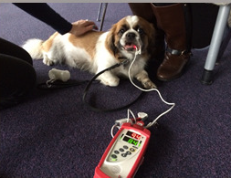 difficulty
eating. Quantitative behavioural observations were conducted on 47
brachycephalic dogs. Behaviour during (i) a 6-minute walk test, (ii) a play
test (iii) and an appetite test was recorded. BOAS severity was measured
using a previously validated Owner Reported Breathing (ORB) score: 0 =
unaffected; 40 = maximum severity. Increasing BOAS severity tended
(non-significantly) to decrease oxygenation after exercise [at right, a
CKCS is being measured with a pulse a oximeter after exercise],
increased time to ingest per food pellet , and decrease in energetic play
behaviours e.g. Play Bow; and increase breathing noises in all three
domains. An owner questionnaire conducted on 2265 brachycephalic dogs
[including cavalier King Charles spaniels] indicated the
severity scores for mild BOAS are 8-15, moderate BOAS are 16-26 and severe
are 27-40 ORB score, with owner-perceived canine welfare significantly
declining as severity increased. Mild BOAS has less effect on canine welfare
than sometimes assumed. However, moderate BOAS affects at least two of the
welfare domains tested and severe BOAS is recognised to negatively impacts
all exercise, appetite and play domains. This information would will help
make recommendations for owners, breeders and veterinarians on appropriate
treatment to maximise welfare in affected dogs.
difficulty
eating. Quantitative behavioural observations were conducted on 47
brachycephalic dogs. Behaviour during (i) a 6-minute walk test, (ii) a play
test (iii) and an appetite test was recorded. BOAS severity was measured
using a previously validated Owner Reported Breathing (ORB) score: 0 =
unaffected; 40 = maximum severity. Increasing BOAS severity tended
(non-significantly) to decrease oxygenation after exercise [at right, a
CKCS is being measured with a pulse a oximeter after exercise],
increased time to ingest per food pellet , and decrease in energetic play
behaviours e.g. Play Bow; and increase breathing noises in all three
domains. An owner questionnaire conducted on 2265 brachycephalic dogs
[including cavalier King Charles spaniels] indicated the
severity scores for mild BOAS are 8-15, moderate BOAS are 16-26 and severe
are 27-40 ORB score, with owner-perceived canine welfare significantly
declining as severity increased. Mild BOAS has less effect on canine welfare
than sometimes assumed. However, moderate BOAS affects at least two of the
welfare domains tested and severe BOAS is recognised to negatively impacts
all exercise, appetite and play domains. This information would will help
make recommendations for owners, breeders and veterinarians on appropriate
treatment to maximise welfare in affected dogs.
Epidemiological associations between brachycephaly and upper respiratory tract disorders in dogs attending veterinary practices in England. Dan G. O’Neill, Caitlin Jackson, Jonathan H. Guy, David B. Church, Paul D. McGreevy, Peter C. Thomson, Dave C. Brodbelt. Canine Genetics & Epidemiology. July 2015;2:10. Quote: "Background: Brachycephalic dog breeds are increasingly common. Canine brachycephaly has been associated with upper respiratory tract (URT) disorders but reliable prevalence data remain lacking. Using primary-care veterinary clinical data, this study aimed to report the prevalence and breed-type risk factors for URT disorders in dogs. Results: The sampling frame included 170,812 dogs attending 96 primary-care veterinary clinics participating within the VetCompass Programme. Two hundred dogs were randomly selected from each of three extreme brachycephalic breed types (Bulldog, French Bulldog and Pug) and three common small-to medium sized breed types (moderate brachycephalic: Yorkshire Terrier and non-brachycephalic: Border Terrier and West Highland White Terrier). Information on all URT disorders recorded was extracted from individual patient records. Disorder prevalence was compared between groups using the chi-squared test or Fisher’s test, as appropriate. During the study, 83 (6.9 %) study dogs died. Extreme brachycephalic dogs (median longevity: 8.6 years, IQR: 2.4-10.8) were significantly younger at death than the moderate and non-brachycephalic group of dogs (median 12.7 years, IQR 11.1-15.0) (P < 0.001). A higher proportion of deaths in extreme brachycephalic breed types were associated with URT disorders (4/24 deaths, 16.7 %) compared with the moderate and non-brachycephalic group (0/59 deaths, 0.0 %) (P = 0.001). The prevalence of having at least one URT disorder in the extreme brachycephalic group was higher (22.0 %, 95 % confidence interval (CI): 18.0-26.0) than in the moderate and non-brachycephalic group (9.7 %, 95 % CI: 7.1-12.3, P < 0.001). The prevalence of URT disorders varied significantly by breed type: Bulldogs 19.5 %, French Bulldogs 20.0 %, Pugs 26.5 %, Border Terriers 9.0 %, West Highland White Terriers 7.0 % and Yorkshire Terriers 13.0 % (P < 0.001). After accounting for the effects of age, bodyweight, sex, neutering and insurance, extreme brachycephalic dogs had 3.5 times (95 % CI: 2.4-5.0, P < 0.001) the odds of at least one URT disorder compared with the moderate and non-brachycephalic group. Conclusions: In summary, this study reports that URT disorders are commonly diagnosed in Bulldog, French Bulldog, Pug, Border Terrier, WHWT and Yorkshire Terrier dogs attending primary-care veterinary practices in England. The three extreme brachycephalic breed types (Bulldog, French Bulldog and Pug) were relatively short-lived and predisposed to URT disorders compared with three other small-to-medium size breed types that are commonly owned (moderate brachycephalic Yorkshire Terrier and non-brachycephalic: Border Terrier and WHWT)."
Brachycephalic Airway Syndrome: Tips for Successful Diagnosis and Surgery. Katrin Saile. DVM360. September 2015. Quote: "Brachycephalic syndrome refers to a combination of abnormalities in the nose, mouth and trachea of certain breeds of dogs that can cause significant respiratory abnormalities. ... Brachycephalic dogs and cats have early ankylosis in the cartilages at the base of their skull, leading to a shortened longitudinal skull axis. The most common breeds affected by brachycephalic syndrome include English bulldogs, French bulldogs, Pugs, and Boston terriers. Cavalier King Charles spaniels, Shih-Tzus, and Pekinese are also over-represented. ... Most commonly, patients present with stenotic nares, an elongated soft palate, and everted laryngeal saccules. Several components of brachycephalic syndrome can be surgically corrected and dogs can have a good prognosis if the disease is not too advanced."
Clinical Features and Outcome of Dogs with Epiglottic Retroversion With or Without Surgical Treatment: 24 Cases. S.C. Skerrett, J.K. McClaran, P.R. Fox, D. Palma. J. Vet. Int. Med. October 2015. Quote: "Background: Published information describing the clinical features and outcome for dogs with epiglottic retroversion (ER) is limited. Hypothesis/Objectives: To describe clinical features, comorbidities, outcome of surgical versus medical treatment and long-term follow-up for dogs with ER. We hypothesized that dogs with ER would have upper airway comorbidities and that surgical management (epiglottopexy or subtotal epiglottectomy) would improve long-term outcome compared to medical management alone. Animals: Twenty-four client-owned dogs [one cavalier King Charles spaniel]. Methods: Retrospective review of medical records to identify dogs with ER that underwent surgical or medical management of ER. Results: Dogs with ER commonly were middle-aged to older, small breed, spayed females with body condition score (BCS) ≥6/9. Stridor and dyspnea were the most common presenting signs. Concurrent or historical upper airway disorders were documented in 79.1% of cases. At last evaluation, 52.6% of dogs that underwent surgical management, and 60% of dogs that received medical management alone, had decreased severity of presenting clinical signs. In dogs that underwent surgical management for ER, the incidence of respiratory crisis decreased from 62.5% before surgery to 25% after surgical treatment. The overall calculated Kaplan–Meier median survival time was 875 days. Conclusion and clinical importance: Our study indicated that a long-term survival of at least 2 years can be expected in dogs diagnosed with epiglottic retroversion. The necessity of surgical management cannot be determined based on this data, but dogs with no concurrent upper airway disorders may benefit from a permanent epiglottopexy to alleviate negative inspiratory pressures."
Impact of Facial Conformation on Canine Health: Brachycephalic Obstructive Airway Syndrome. Rowena M. A. Packer, Anke Hendricks, Michael S. Tivers, Charlotte C. Burn. PlosOne. October 2015. Quote: "The domestic dog may be the most morphologically diverse terrestrial mammalian species known to man; pedigree dogs are artificially selected for extreme aesthetics dictated by formal Breed Standards, and breed-related disorders linked to conformation are ubiquitous and diverse. Brachycephaly–foreshortening of the facial skeleton–is a discrete mutation that has been selected for in many popular dog breeds e.g. the Bulldog, Pug, and French Bulldog. A chronic, debilitating respiratory syndrome, whereby soft tissue blocks the airways, predominantly affects dogs with this conformation, and thus is labelled Brachycephalic Obstructive Airway Syndrome (BOAS). Despite the name of the syndrome, scientific evidence quantitatively linking brachycephaly with BOAS is lacking, but it could aid efforts to select for healthier conformations. Here we show, in (1) an exploratory study of 700 dogs of diverse breeds and conformations [including 26 cavalier King Charles spaniels], and (2) a confirmatory study of 154 brachycephalic dogs [including 11 CKCSs], that BOAS risk increases sharply in a non-linear manner as relative muzzle length shortens. BOAS only occurred in dogs whose muzzles comprised less than half their cranial lengths. Thicker neck girths also increased BOAS risk in both populations: a risk factor for human sleep apnoea and not previously realised in dogs; and obesity was found to further increase BOAS risk. This study provides evidence that breeding for brachycephaly leads to an increased risk of BOAS in dogs, with risk increasing as the morphology becomes more exaggerated. As such, dog breeders and buyers should be aware of this risk when selecting dogs, and breeding organisations should actively discourage exaggeration of this high-risk conformation in breed standards and the show ring."
Clinical effects of the use of a bipolar vessel sealing device for soft palate resection and tonsillectomy in dogs, with histological assessment of resected tonsillar tissue. DA Cook, PA Moses, JT Mackie. Australian Vet. J. November 2015;93(12):445-451. Quote: "Objective: To investigate whether soft palate resection and tonsillectomy with a bipolar vessel sealing device (BVSD) improves clinical respiratory score. To document histopathological changes to tonsillar tissue following removal with a BVSD. Methods & Results: Case series of 22 dogs with clinical signs of upper respiratory obstruction related to brachycephalic airway syndrome. Soft palate and tonsils were removed using a BVSD. Alarplasty and saccullectomy were also performed if indicated. A clinical respiratory score was assigned preoperatively, 24-h postoperatively and 5 weeks postoperatively. Excised tonsillar samples were measured and then assessed histologically for depth of tissue damage deemed to be caused by the device. Depth of tissue damage was compared between two power settings of the device. Soft palate resection and tonsillectomy with a BVSD lead to a significant improvement in respiratory scores following surgery. Depth of tissue damage was significantly less for power setting 1 compared with power setting 2. Using power setting 1, median calculated depth of tonsillar tissue damage was 3.4 mm (range 1.2–8.0). One dog experienced major complications. Conclusion: Soft palate resection and tonsillectomy with a BVSD led to significant improvement in clinical respiratory score."
RETURN TO TOP
2016
Histopathologic and immunohistochemical features of soft palate muscles and nerves in dogs with an elongated soft palate. Kiyotaka Arai, Masanori Kobayashi, Yasuji Harada, Yasushi Hara, Masaki Michishita, Kozo Ohkusu-Tsukada, Kimimasa Takahashi. Amer. J. Vet. Research. January 2016;77(1):77-83. Quote: "Objective: To histologically evaluate and compare features of myofibers within the elongated soft palate (ESP) of brachycephalic and mesocephalic dogs with those in the soft palate of healthy dogs and to assess whether denervation or muscular dystrophy is associated with soft palate elongation. Sample: Soft palate specimens from 24 dogs with ESPs (obtained during surgical intervention) and from 14 healthy Beagles (control group). ... The brachycephalic breeds included French Bulldog (n = 6), Pug (5), Pomeranian (3), Shih Tzu (2), Cavalier King Charles Spaniel (2), Pekinese (1), and Bulldog (1). ... Procedures: All the soft palate specimens underwent histologic examination to assess myofiber atrophy, hypertrophy, hyalinization, and regeneration. The degrees of atrophy and hypertrophy were quantified on the basis of the coefficient of variation and the number of myofibers with hyalinization and regeneration. The specimens also underwent immunohistochemical analysis with anti-neurofilament or anti-dystrophin antibody to confirm the distribution of peripheral nerve branches innervating the palatine myofibers and myofiber dystrophin expression, respectively. Results: Myofiber atrophy, hypertrophy, hyalinization, and regeneration were identified in almost all the ESP specimens. Degrees of atrophy and hypertrophy were significantly greater in the ESP specimens, compared with the control specimens. There were fewer palatine peripheral nerve branches in the ESP specimens than in the control specimens. Almost all the myofibers in the ESP and control specimens were dystrophin positive. Conclusions and Clinical Relevance: These results suggested that palatine myopathy in dogs may be caused, at least in part, by denervation of the palatine muscles and not by Duchenne- or Becker-type muscular dystrophy. These soft palate changes may contribute to upper airway collapse and the progression of brachycephalic airway obstructive syndrome."
Canine tracheal collapse. S. W. Tappin. J. Sm. Anim. Pract. January 2016;57(1):9-17. Quote: "Tracheal collapse occurs most commonly in middle-aged, small breed dogs. Clinical signs are usually proportional to the degree of collapse, ranging from mild airway irritation and paroxysmal coughing to respiratory distress and dyspnoea. Diagnosis is made by documenting dynamic airway collapse with radiographs, bronchoscopy or fluoroscopy. Most dogs respond well to medical management and treatment of any concurrent comorbidities. Surgical intervention may need to be considered in dogs that do not respond or have respiratory compromise. A variety of surgical techniques have been reported although extraluminal ring prostheses or intraluminal stenting are the most commonly used. Both techniques have numerous potential complications and require specialised training and experience but are associated with good short- and long-term outcomes."
Comparison between computed tomographic characteristics of the middle ear in nonbrachycephalic and brachycephalic dogs with obstructive airway syndrome. Raquel Salgüero, Michael Herrtage, Mark Holmes, Paddy Mannion, Jane Ladlow. Vet. Radiology & Ultrasound. March 2016;57(2):137-143. Quote: “Prevalence of subclinical middle ear lesions in dogs that undergo computed tomography (CT) and magnetic resonance imaging of the head has been reported up to 41%. ... Eustachian tube dysfunction has been postulated to be the cause of the high prevalence (54%) of middle ear effusions in Cavalier King Charles spaniels. ... A predisposition in brachycephalics has been suggested, however evidence-based studies are lacking. Aims of this retrospective cross-sectional study were to compare CT characteristics of the middle ear in groups of nonbrachycephalic and brachycephalic dogs that underwent CT of the head for conditions unrelated to ear disease, and test associations between thickness of the soft palate and presence of subclinical middle ear lesions. One observer recorded CT findings for each dog without knowledge of group status. A total of 65 dogs met inclusion criteria (25 brachycephalic, 40 nonbrachycephalic). Brachycephalic dogs had a significantly thicker bulla wall (P = 2.38 × 10⁻²⁶) and smaller luminal volume (P = 5.74 × 10⁻²⁰), when compared to nonbrachycephalic dogs. Soft palate thickness was significantly greater in the brachycephalic group (P = 2.76 × 10⁻⁹). Nine of 25 brachy-cephalic dogs had material in the lumen of the tympanic cavity, compared to zero of 45 of non-brachycephalics. Within the brachycephalic group, a significant difference in mean soft palate thickness was identified for dogs with material in the middle ear (12.2 mm) vs. air-filled bullae (9 mm; P = 0.016). Findings from the current study supported previous theories that brachycephalic dogs have a greater prevalence of subclinical middle ear effusion and smaller bulla luminal size than nonbrachycephalic dogs. ... When the Eustachian tube function is impaired, middle ear pressure becomes more negative, drawing extracellular fluid into the bulla and producing serous effusion. 12 Similar changes were found in Cavalier King Charles spaniels, where a greater thickness of the soft palate and reduced nasopharyngeal aperture were significantly associated with subclinical otitis media ... Authors recommend that the bulla lumen volume formula previously developed for mesaticephalic dogs, (−0.612 + 0.757 [lnBW]) be adjusted to 1/3(−0.612 + 0.757 [lnBW]) for brachycephalic breeds. ... Further prospective studies are needed with a larger number of brachycephalic dogs and to compare our breeds with Cavalier King Charles spaniels as previous publications have already shown the presence of subclinical otitis media in this breed.”
How does multilevel upper airway surgery influence the lives of dogs with severe brachycephaly? Results of a structured pre- and postoperative questionnaire administered to dog owners. S. Pohla, F. Roedlera, G.U. Oechtering. Vet. J. February 2016. Quote: "Using a structured questionnaire, the present study investigated the dog owner-perceived severity and frequency of a broad spectrum of welfare-relevant impairments 2 weeks before and 6 months after brachycephalic dogs underwent a recently developed multi-level upper airway surgery. All dogs underwent surgical treatment of stenotic nares (ala-vestibuloplasty), the nasal cavity (laser-assisted turbinectomy, LATE), the pharynx (palatoplasty and tonsillotomy), and if indicated, laryngeal surgery (laser-assisted ablation of everted ventricles and partial cuneiformectomy). Questionnaire data from owners of 37 Pugs and 25 French bulldogs were evaluated. In all dogs, the clinical signs associated with brachycephaly improved markedly after surgery. Most encouraging was the striking reduction in life-threatening events by 90% (choking fits decreased from 60% to 5% and collapse from 27% to 3%). The incidence of sleeping problems decreased from 55% to 3%, and the occurrence of breathing sounds declined by approximately 50%. There was a marked improvement in exercise tolerance and a modest improvement in heat tolerance. Dogs with severe brachycephaly benefitted substantially from multi-level surgery, and there were particular improvements in the incidences of severe impairment and life-threatening events. However, despite the marked improvement perceived by dog owners, these dogs remained clinically affected and continued to show welfare-relevant impairments caused by these hereditary disorders.
Brachycephalic Syndrome. Gilles Dupré, Dorothee Heidenreich. Vet. Clinics of No. America: Sm. Anim. Pract. March 2016. Quote: "Brachycephalic syndrome (BS) is an established cause of respiratory distress in brachycephalic breeds. Breeds most commonly affected are English and French bulldogs, pugs, and Boston terriers; however, Pekingese, Shih tzu, Cavalier King Charles Spaniels, Boxers, Dogue de Bordeaux, and Bullmastiffs are also categorized as brachycephalic dogs. Most owners report heat, stress and exercise intolerance, snoring, inspiratory dyspnea, and in severe cases, cyanosis and even syncopal episodes. Sleep apneas can be observed,5 and occasionally gastrointestinal signs such as vomiting and regurgitation. KEY POINTS: Skull conformation anomalies in brachycephalic breeds lead to compression of nasal passages. Additional mucosal hyperplasia and secondary collapse of the upper airway contribute to a multilevel obstruction and the genesis of the so-called brachycephalic syndrome. Surgical treatments usually include widening of stenotic nares as well as various palatoplasty techniques to improve airflow through the rima glottidis. The overall prognosis for a significant improvement is excellent."
Efficacy of bronchial stenting in dogs with myxomatous mitral valve disease and bronchial collapse. Dar Ozer, Samantha Siess, Brienne Williams, Nikki Gaudette, George Kramer. J. Vet. Int. Med. June 2016. 2016 ACVIM Forum Abstract C-03. Quote: Chronic airway disease and myxomatous mitral valve disease (MMVD) are frequent comorbidities in small breed dogs. Bronchial collapse can cause coughing, tachypnea and hypoxemia. Pulmonary hypertension can be seen with both MMVD and chronic airway disease. The purpose of this study was to review outcomes in dogs that had bronchial stents placed due to bronchial collapse. It was hypothesized that moderate to severe MMVD or the presence of pulmonary hypertension would not have a negative effect on lifespan after stent placement. Medical records of 18 small breed dogs that had bronchial stent placement for chronic coughing secondary to bronchial collapse were reviewed. A hierarchical multiple linear regression analysis was conducted to predict lifespan after bronchial stent placement based on age at time of stent placement, severity of MMVD and the presence of pulmonary hypertension. Eighteen dogs had bronchial stents placed over a period of 7 years. Age at the time of stent placement ranged from 6.5 years to 14 years of age (M = 10.47 1.85). Breeds represented included Cavalier King Charles spaniel (2), beagle (2), Chihuahua (4), Pomeranian (3), toy poodle (2), Yorkshire terrier (2), Maltese (1), Coton de Tulear (1) and shih tzu (1). There were 11 males and 7 females. Twelve dogs (66.67%) had evidence of moderate to severe MMVD and 4 (22.22%) had evidence of pulmonary hypertension. Six dogs (33.33%) had CHF prior to stent placement and 6 dogs (33.33%) had CHF after stent placement. Syncope was reported in 6 dogs prior to stent placement and 5 dogs after stent placement. The average lifespan after stent placement was 203.56 250.72 days. Three dogs are currently alive post-stent placement (1013, 559 and 411 days). A hierarchical multiple linear regression was calculated to predict lifespan after placement of a bronchial stent based on age at the time of stent placement, the presence of pulmonary hypertension, and severity of MMVD. In stage one, age at the time of stent placement significantly predicted lifespan after placement of a bronchial stent (b = 7.10, P < .02); with lifespan decreasing by 7.10 days for each additional month of age at the time of stent placement. The presence of pulmonary hypertension did not significantly predict lifespan (b = 235.32, P = .10). Severity of MMVD did not contribute significantly to the model in stage two (b = 101.38, P = 0.43). Results from this study indicate that the severity of MMVD or the presence of pulmonary hypertension did not negatively affect lifespan after bronchial stent placement. As such, moderate to severe MMVD and the presence of pulmonary hypertension should not be viewed as exclusion criteria when assessing candidates for possible bronchial stenting. Prospective studies should be conducted to further investigate the clinical benefit of bronchial stenting in dogs with severe bronchial collapse.
Brachycephalic Syndrome. Comparison Between Conventional Surgical Techniques, Resection with Surgical Laser and Plasty. Advantages and Disadvantages of Each. Gala Secanella Garcés. Univ. Barcelona. June 2016. Quote: Dog breeds most commonly affected are English and French bulldogs, Pugs, Boston terriers, Pekingese, Shih tzu, Maltese, Cavalier King Charles Spaniels, Boxers, Yorkshire terriers, Pinschers miniature, Dogue de Bordeaux and Bullmastiffs. ... The definitive treatment for the BS [brachycephalic syndrome] is always surgical. Early intervention can slow down the progression of the signs and complications. There are several surgical techniques and, during the last years, the use of CO2 laser ensures less surgical time, bleeding, swelling and intra and postoperative pain, as well as a more precise tissue ressection. A possible explanation for the poor therapeutic success after conventional surgeries, could be the lack of consideration in the diagnosis, management and treatment of the rest of intranasal structures. To understand the BS, all the efforts should relapse into the selection of these breeds, so that in the future, the objective is to encourage more moderate craniofacial morphologies in order to reduce the prevalence and severity of BS.
Use of CT to evaluate and compare intranasal features in brachycephalic
and normocephalic dogs. M. Auger, K. Alexander, G. Beauchamp, M. Dunn.
J. Small Animal Pract. October 2016;57(10):529-536.
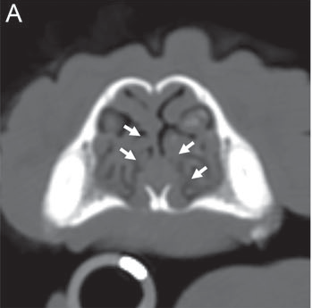 Quote:
Objectives: To evaluate and compare nasal mucosal contact, septal deviation
and caudal aberrant nasal turbinates in brachycephalic and normocephalic
dogs using computed tomography. Methods: Dogs without nasal disease and
having undergone computed tomography scan of the head (plica alaris to the
cribiform plate) were retrospectively selected and divided into
brachycephalic and normocephalic groups. Eighteen brachycephalic [including
two cavalier King Charles spaniels] and 32 normocephalic
dogs were included. Anatomic criteria were used to locate predetermined
pairs of intranasal structures and nasal mucosal contact was described as
present or absent for each site. Septal deviations were identified and
measured using angle of septal deviation. Caudal aberrant nasal turbinates
were identified and categorised when present. [View at right shows
contact between the concha nasalis ventralis and the nasal septum in a 7.3
year old CKCS.] Results: Prevalence of nasal mucosal
contact was significantly higher in brachycephalic dogs. No significant
difference was seen in prevalence or in angle of septal deviation between
groups. Prevalence of caudal aberrant nasal turbinates was significantly
higher in brachycephalic dogs. Clinical Significance: Nasal mucosal contact
and caudal aberrant nasal turbinates were significantly more prevalent in
brachycephalic dogs than in normocephalic dogs in our study. Computed
tomography can be a valuable aid in obtaining data on nasal mucosal contact,
caudal aberrant nasal turbinates and septal deviations. Combination of
computed tomography with endoscopy and functional airway testing would be
useful to further evaluate the correlation between intranasal features and
symptoms of brachycephalic airway syndrome.
Quote:
Objectives: To evaluate and compare nasal mucosal contact, septal deviation
and caudal aberrant nasal turbinates in brachycephalic and normocephalic
dogs using computed tomography. Methods: Dogs without nasal disease and
having undergone computed tomography scan of the head (plica alaris to the
cribiform plate) were retrospectively selected and divided into
brachycephalic and normocephalic groups. Eighteen brachycephalic [including
two cavalier King Charles spaniels] and 32 normocephalic
dogs were included. Anatomic criteria were used to locate predetermined
pairs of intranasal structures and nasal mucosal contact was described as
present or absent for each site. Septal deviations were identified and
measured using angle of septal deviation. Caudal aberrant nasal turbinates
were identified and categorised when present. [View at right shows
contact between the concha nasalis ventralis and the nasal septum in a 7.3
year old CKCS.] Results: Prevalence of nasal mucosal
contact was significantly higher in brachycephalic dogs. No significant
difference was seen in prevalence or in angle of septal deviation between
groups. Prevalence of caudal aberrant nasal turbinates was significantly
higher in brachycephalic dogs. Clinical Significance: Nasal mucosal contact
and caudal aberrant nasal turbinates were significantly more prevalent in
brachycephalic dogs than in normocephalic dogs in our study. Computed
tomography can be a valuable aid in obtaining data on nasal mucosal contact,
caudal aberrant nasal turbinates and septal deviations. Combination of
computed tomography with endoscopy and functional airway testing would be
useful to further evaluate the correlation between intranasal features and
symptoms of brachycephalic airway syndrome.
Perfusion-weighted Magnetic Resonance Imaging of Brachycephalic Dogs with Ventriculomegaly. M. J. Schmidt, A. Hartmann, K. H. von Pückler, L. Hähnsen, N. Ondreka. Vet. Rad. & Ultra. October 2016;57(6):646-669. Quote: Introduction: It has been shown that ventriculomegaly in dogs is associated with a loss of periventricular white matter comparable to clinical relevant internal hydrocephalus or normal pressure hydrocephalus (NPH). The pathophysiology of ventricular distension is unclear currently. The aim of this study was to compare brain perfusion in dogs with and without ventriculomegaly using perfusion-weighted magnetic resonance imaging to clarify, as to whether abnormal perfusion might be involved in the pathophysiology of ventriculomegaly. Materials and methods: Perfusion was measured in 23 Cavalier King Charles spaniels with ventriculomegaly, 10 healthy Beagle dogs were examined as a control group. Perfusion-weighted images were acquired using a dynamic multishot fast-field echo-EPI in a dorsal plane. Forty acquisitions per slice were acquired. Contrast was automatically injected using an automatic pump (0.2 mmol/kg body weight) in 20 ml crystalloid solution with an injection rate of 5 ml/s. Contrast was given at the 10th acquisition. Perfusion was measured using free-hand regions of interest (ROI) in five brain regions: periventricular white matter, caudate nucleus, parietal cortex, hippocampus, and thalamus using the STROKETOOL software tool. Time to peak (TTP), regional cerebral blood flow (rCBF), regional blood volume (rCBV), and mean transit time (MTT) was measured. Intraobserver variability was measured using kappa statistics. Differences between groups were tested using Mann–Whitney U-tests. Significance level was <0.05. Results: rCBF and rCBV were significantly lower in the periventricular white matter of the dogs with ventriculomegaly compared to control dogs (P < 0.001) but not for the other ROIs. Kappa statistics revealed substantial agreement between measurements (0.82). Discussion: Based on the differences in periventricular perfusion ventriculomegaly should be interpreted as a form of NPH as suggested previously. The impaired perfusion might be the reason for the reduced white matter volume in these dogs. This has been shown in laboratory rodents with experimentally induced hydrocephalus and humans with NPH. Cognitive studies should be conducted to investigate a possible impairment of memory, learning, and spatial orientation as present in humans with NPH.
Developmental and Evolutionary Significance of the Zygomatic Bone. Yann Heuzé, Kazuhiko Kawasaki, Tobias Schwarz, Jeffrey J. Schoenebeck, Joan T. Richtsmeier. Anat. Rec. December 2016;299(12):1616-1630. Quote: The zygomatic bone is derived evolutionarily from the orbital series. In most modern mammals the zygomatic bone forms a large part of the face and usually serves as a bridge that connects the facial skeleton to the neurocranium. Our aim is to provide information on the contribution of the zygomatic bone to variation in midfacial protrusion using three samples; humans, domesticated dogs, and monkeys. In each case, variation in midface protrusion is a heritable trait produced by one of three classes of transmission: localized dysmorphology associated with single gene dysfunction, selective breeding, or long-term evolution from a common ancestor. We hypothesize that the shape of the zygomatic bone reflects its role in stabilizing the connection between facial skeleton and neurocranium and consequently, changes in facial protrusion are more strongly reflected by the maxilla and premaxilla. Our geometric morphometric analyses support our hypothesis suggesting that the shape of the zygomatic bone has less to do with facial protrusion. By morphometrically dissecting the zygomatic bone we have determined a degree of modularity among parts of the midfacial skeleton suggesting that these components have the ability to vary independently and thus can evolve differentially. From these purely morphometric data, we propose that the neural crest cells that are fated to contribute to the zygomatic bone experience developmental cues that distinguish them from the maxilla and premaxilla. The spatiotemporal and molecular identity of the cues that impart zygoma progenitors with their identity remains an open question that will require alternative data sets. ... When the shape of the left canine facial skeleton is analyzed using PCA [principal components analysis], the first PC accounts for 79% of the total shape variation for which size (CS) explains 57% (P < 0.0001). Shape variation of the canine facial skeleton separates canine individuals primarily along PC1 (Fig. 6a) according to the relative magnitude of facial retrusion with the pug and bulldog, and to a lesser degree Lhasa apso, cavalier King Charles spaniel, and boxer tending towards the negative end of PC1 (retrognathic dogs), and the Scottish terrier, flat coated retriever and greyhound anchoring the positive end (prognathic dogs). ... Given the variation in size among dog breeds, we were interested in the relative size variation of the different anatomical units of the facial skeleton studied. To determine whether size varies in a pattern similar among the different anatomical units we studied (i.e., facial skeleton, premaxilla, and maxilla, zygoma), we used centroid size of the facial skeleton, premaxilla, maxilla, and zygoma as a proxy for size of each anatomical unit. The mean plot of these four anatomical units grouped by breed indicates that size varies according to a similar pattern among the different anatomical units forming the facial skeleton. The zygoma however appears a bit “out of phase” (larger than expected) relative to the pattern of centroid size estimated for the other facial units for the retrognathic breeds (boxer, bulldog, pug and Rottweiler) and the cavalier King Charles spaniel and cocker spaniel.
RETURN TO TOP
2017
Use of Morphometric Mapping to Characterise Symptomatic Chiari-Like
Malformation, Secondary Syringomyelia and Associated Brachycephaly in the
Cavalier King Charles Spaniel. Susan P. Knowler,
Chloe Cross, Sandra Griffiths, Angus K. McFadyen, Jelena Jovanovik, Anna
Tauro, Zoha Kibar, Colin J. Driver, Roberto M. La Ragione, Clare Rusbridge.
PLOS One. January 2017. Quote: Objectives: To characterise the symptomatic
phenotype of Chiari-like malformation (CM), secondary syringomyelia (SM) and
brachycephaly in the Cavalier King Charles Spaniel [CKCS]
using morphometric [the quantitative analysis of form, a concept that
encompasses size and shape] measurements on mid-sagittal Magnetic Resonance
images (MRI) of the brain and craniocervical junction. Methods: This
retrospective study, based on a previous quantitative analysis in the
Griffon Bruxellois (GB), used 24 measurements taken on 130 T1-weighted MRI
of hindbrain and cervical region. Associated brachycephaly was estimated
using 26 measurements, including rostral forebrain flattening and olfactory
lobe rotation, on 72 T2-weighted MRI of the whole brain. Both study cohorts
were divided into three groups; Control, CM pain and SM and their
morphometries compared with each other. Results: Fourteen significant traits
were identified in the hindbrain study and nine traits in the whole brain
study, six of which were similar to the GB and suggest a common aetiology.
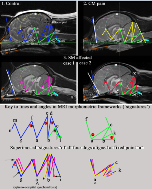 The
Control cohort [31 CKCS in the hindbrain study (HBS) and 16
CKCSs in the whole brain study (WBS)] had the most natural,
wolf-like, skull conformation in terms of ellipticity. The CM pain cohort
[28 CKCSs in HBS and 25 CKCSs in WBS] was
characterised by increased brachycephaly with greatest rostral forebrain
flattening, shortest basicranium and compensatory cranial height. However,
in this cohort, an increased distance between the occiput and atlas provided
fewer impediments to CSF dynamics at the foramen magnum and reduced the risk
for SM. The SM cohort [71 CKCSs in HBS and 31 CKCSs
in WBS] exhibited two conformation anomalies. One phenotype variation was
influenced by incongruities at the craniocervical junction and increased
proximity of the dens producing a ‘concertina’ type flexure with medullary
elevation. The other phenotypic variation was influenced by increased
brachycephaly resulted in a ‘concertina’ type flexure similar to the CM pain
cohort. However, both SM variations were characterised by an apparent
reduction in caudal fossa volume which compromised the CSF dynamics in the
spinal cord. Conclusion: Morphometric mapping provides a diagnostic tool for
quantifying symptomatic CM, secondary SM and their relationship with
brachycephaly. It might identify dogs at risk of SM and CM pain to improve
diagnosis and make available a means for screening breeding dogs and provide
estimated breeding values. It is hypothesized that CM pain is associated
with increased brachycephaly and SM can result from different combinations
of abnormalities of the forebrain, caudal fossa and craniocervical junction
which compromise the neural parenchyma and impede cerebrospinal fluid flow.
(Motion
picture highlights the dynamic changes of the skull conformation and
brain parenchyma associated with progressive brachycephaly and airorhynchy,
shortening of the basicranium and supraoccipital bones and the proximity and
angulation of the atlas and dens.)
The
Control cohort [31 CKCS in the hindbrain study (HBS) and 16
CKCSs in the whole brain study (WBS)] had the most natural,
wolf-like, skull conformation in terms of ellipticity. The CM pain cohort
[28 CKCSs in HBS and 25 CKCSs in WBS] was
characterised by increased brachycephaly with greatest rostral forebrain
flattening, shortest basicranium and compensatory cranial height. However,
in this cohort, an increased distance between the occiput and atlas provided
fewer impediments to CSF dynamics at the foramen magnum and reduced the risk
for SM. The SM cohort [71 CKCSs in HBS and 31 CKCSs
in WBS] exhibited two conformation anomalies. One phenotype variation was
influenced by incongruities at the craniocervical junction and increased
proximity of the dens producing a ‘concertina’ type flexure with medullary
elevation. The other phenotypic variation was influenced by increased
brachycephaly resulted in a ‘concertina’ type flexure similar to the CM pain
cohort. However, both SM variations were characterised by an apparent
reduction in caudal fossa volume which compromised the CSF dynamics in the
spinal cord. Conclusion: Morphometric mapping provides a diagnostic tool for
quantifying symptomatic CM, secondary SM and their relationship with
brachycephaly. It might identify dogs at risk of SM and CM pain to improve
diagnosis and make available a means for screening breeding dogs and provide
estimated breeding values. It is hypothesized that CM pain is associated
with increased brachycephaly and SM can result from different combinations
of abnormalities of the forebrain, caudal fossa and craniocervical junction
which compromise the neural parenchyma and impede cerebrospinal fluid flow.
(Motion
picture highlights the dynamic changes of the skull conformation and
brain parenchyma associated with progressive brachycephaly and airorhynchy,
shortening of the basicranium and supraoccipital bones and the proximity and
angulation of the atlas and dens.)
Time-dependent changes and prognostic value of lactatemia during the first 24 hours of life in brachycephalic newborn dogs. C. Castagnetti, M. Cunto, C. Bini, J. Mariella, S. Capolongo, D. Zam. Theriogenology. February 2017. Quote: Blood lactate concentration is known to be a good prognostic indicator associated with the severity of illness and the patient's outcome both in human and veterinary medicine. It also plays a significant role in the assessment of the newborn, being a good indicator of fetal hypoxia and the ideal predictor of morbidity at term in babies. In veterinary neonatal medicine, hyperlactatemia is considered a valid prognostic marker in critically ill foals; moreover, blood lactate measurement has been proposed for the evaluation of newborn viability and the assessment of fetal distress during delivery in dogs. Unfortunately, only a few studies have been published concerning the canine species. The present work examines 67 brachycephalic newborn dogs and their mothers, with the aim to evaluate the time-dependent changes of blood lactate and glucose concentration during the first 24 h after vaginal or caesarean delivery both in puppies and bitches. ... The study included seven pregnant bitches examined in the period 2012-2014. Six of them were English Bulldogs (four belonging to a breeding kennel and two private-owned) and one was a private-owned Cavalier King Charles Spaniel. ... To our knowledge, this is the first published study examining the time-dependent changes of these parameters in the bitch after parturition. Within the studied population of puppies, non-surviving was significantly associated with a higher lactatemia and a lower APGAR score. Blood lactate was high at birth then progressively decreased during the first 24 h of life and a lack of normalization of blood lactate levels within this time interval was suggestive for a poor prognosis for the newborn dogs; moreover, the decrease appeared to be slower after vaginal delivery. Lactatemia also showed a positive correlation with glycemia at birth. Concerning the bitches examined, blood lactate was found to be significantly higher after vaginal delivery than after caesarean section; the normalization occurred within 24 h after parturition. Blood glucose level was significantly higher at 2 h from delivery both in the group of bitches submitted to caesarean section and in those undergoing natural whelping but no statistical correlation was found between maternal glycemia and lactatemia. The results of the present study highlighted that the monitoring of lactatemia during the first 24 h of life, in association with the assessment of the APGAR score at birth, can be an useful prognostic tool helping to identify the most severely distressed puppies and to provide them an adequate support. Hghlights: • Lactate plays a significant role in the assessment of the newborn, both in human and veterinary medicine. • There are no published studies reporting the time-dependent changes of lactatemia during the first 24 h of life either in brachycephalic dogs or in other breeds. • The clearance of blood lactate is more rapid after caesarean section, since in this case puppies do not experience the stress of labour. • The time-dependent decrease of blood lactate concentration is missing or very slow in non-surviving puppies. • Monitoring of lactatemia during the first 24 h life, in association with the assessment of the APGAR score at birth, can be an useful prognostic tool helping to identify the most severely distressed puppies.
Anesthetic Protocols for Brachycephalic Dogs. Tasha McNerney. Vet. Team Brief. March 2017. Quote: Brachycephalic dogs are becoming more popular as pets, which means veterinary nurses are more likely to be asked to anesthetize these dogs in practice. Brachycephalic dogs have a relatively broad, short skull, usually with the breadth at least 80% of the length.2 They often have anatomic abnormalities (eg, stenotic nares, elongated soft palate, hypoplastic trachea, laryngeal collapse, everted laryngeal saccules), known as brachycephalic syndrome, which can cause upper airway obstruction and mandate the use of special protocols when administering anesthesia.
The Nationwide Brachycephalic Breed Disease Prevalence Study: Short-nosed breeds more often affected by common conditions, not just known issues. NationwideDVM.com March 2017. Quote: The American Kennel Club lists three brachycephalic breeds (bulldogs, French bulldogs and boxers) in its top 10 most popular breeds; seven in the organization’s top 25. The AKC reports the French bulldog is the most popular breed in New York City, with bulldogs and Cavalier King Charles spaniels at No. 1 in select Manhattan ZIP codes. Nationwide’s proprietary claims data bears out the popularity of these breeds. Of the 1.27 million dogs whose health insurance claims were analyzed for this study [Figure 1], 15% were members of one of the 24 breeds identified as brachycephalic [including the CKCS]. (Nationwide’s list of all breeds includes more than 300 purebreds and crossbreds, not including five classifications of mixed breeds by size.) ... By examining the issues of brachycephalic dogs from a different angle—analyzing common conditions, not brachycephalic-specific ones—Nationwide has expanded the field of inquiry and set a foundation for further study. In so doing, the company is assisting veterinarians, pet owners, breed associations and other stakeholders in the common goal of working together to improve the health of these popular dogs. With an analysis that shows brachycephalic breeds significantly more impacted than their structurally normal counterparts across a range of common conditions, Nationwide has opened a new topic for discussion. In so doing, we hope to help improve the quality of life for these dogs, and to lessen the expense their owners have in caring for them.
Canine Brachycephaly Is Associated with a Retrotransposon-Mediated Missplicing of SMOC2. Thomas W. Marchant, Edward J. Johnson, Lynn McTeir, Craig I. Johnson, Adam Gow, Tiziana Liuti, Dana Kuehn, Karen Svenson, Mairead L. Bermingham, Michaela Drögemüller, Marc Nussbaumer, Megan G. Davey, David J. Argyle, Roger M. Powell, Sérgio Guilherme, Johann Lang, Gert Ter Haar, Tosso Leeb, Tobias Schwarz, Richard J. Mellanby, Dylan N. Clements, Jeffrey J. Schoenebeck. Current Biology. May 2017. Quote: In morphological terms, “form” is used to describe an object’s shape and size. In dogs, facial form is stunningly diverse. Facial retrusion, the proximodistal shortening of the snout and widening of the hard palate is common to brachycephalic dogs and is a welfare concern, as the incidence of respiratory distress and ocular trauma observed in this class of dogs is highly correlated with their skull form. Progress to identify the molecular underpinnings of facial retrusion is limited to association of a missense mutation in BMP3 among small brachycephalic dogs. Here, we used morphometrics of skull isosurfaces derived from 374 pedigree and mixed-breed dogs to dissect the genetics of skull form [including 6 cavalier King Charles spaniels]. Through deconvolution of facial forms, we identified quantitative trait loci that are responsible for canine facial shapes and sizes. Our novel insights include recognition that the FGF4 retrogene insertion, previously associated with appendicular chondrodysplasia, also reduces neurocranium size. Focusing on facial shape, we resolved a quantitative trait locus on canine chromosome 1 to a 188-kb critical interval that encompasses SMOC2. An intronic, transposable element within SMOC2 promotes the utilization of cryptic splice sites, causing its incorporation into transcripts, and drastically reduces SMOC2 gene expression in brachycephalic dogs. SMOC2 disruption affects the facial skeleton in a dose-dependent manner. The size effects of the associated SMOC2 haplotype are profound, accounting for 36% of facial length variation in the dogs we tested. Our data bring new focus to SMOC2 by highlighting its clinical implications in both human and veterinary medicine.
Postanaesthetic pulmonary oedema in a dog following intravenous naloxone administration after upper airway surgery. Natalie Bruniges, Clara Rigotti. Vet. Rec. Casereports. June 2017;5(2). Quote: A cavalier King Charles spaniel was anaesthetised for upper airway surgery. A constant rate infusion of fentanyl at 6 μg/kg/hour and top-up boluses (5 μg/kg in total) were used for intraoperative analgesia. Intermittent positive pressure ventilation (IPPV) was instituted due to tachypnoea and inability to maintain normocapnia. Apnoea and severe hypercapnia developed after cessation of IPPV. IPPV was recommenced for 10 min to reduce hypercapnia, after which spontaneous ventilation returned. The patient had not awakened 45 minutes after isoflurane was turned off and 0.01 mg/kg naloxone was administered intravenously due to suspected fentanyl-induced narcosis. Following immediate arousal, the patient vomited and suddenly developed symptoms and radiographic changes consistent with pulmonary oedema. General anaesthesia was reinduced and 1 mg/kg furosemide was administered intravenously. IPPV was started with application of positive end expiratory pressure in an air/oxygen mixture for 60 minutes. Recovery was uneventful. This is the first report of a dog developing pulmonary oedema following intravenous naloxone.
Elongated Soft Palate Resection with a Flexible Fiber CO2 Laser. Ray Arza. Clinician's Brief. October 2017;15(10):84.
Complications following laryngeal sacculectomy in brachycephalic dogs. J. R. Hughes, B. M. Kaye, A. R. Beswick, G. Ter Haar. J. Sm. Anim. Pract. October 2017. DOI:10.1111/jsap.12763. Quote: Objectives: To evaluate the effect of sacculectomy on the immediate postoperative complication rate in dogs affected with brachycephalic obstructive airway syndrome. Materials and Methods: Retrospective review of clinical records of brachycephalic dogs [limited to pugs, British bulldogs, and French bulldogs] with everted saccules that underwent surgery for brachycephalic obstructive airway syndrome between 2009 and 2014. Dogs were grouped as those having nares resection and staphylectomy only and those having nares resection, staphylectomy and laryngeal sacculectomy. Complications were scored as mild, moderate or severe. Results: In total, 37 dogs were included in the sacculectomy group and 44 in the comparator group. Dogs that had undergone sacculectomy were more likely to develop postoperative complications, with 18 of 37 developing complications, nine of which were moderate to severe. In the group without sacculectomy, nine of 44 dogs developed complications, of which one was severe. Different breed distribution between groups might also impact this outcome. Clinical Significance: The results suggest that sacculectomy might increase morbidity following brachycephalic airway surgery, but repeat studies are required to confirm this result. Further information is also required to determine whether the short-term risks of sacculectomy are outweighed by superior long-term functional outcome.
Anatomical features of the optic canal and the cephalic index in the cranial bone of healthy dogs. Y Ichikawa, N Kanemaki. Vet. Ophthal. October 2017. DOI: 10.1111/vop.12519; E-14. Quote: Purpose: We studied the anatomical features of the optical canal among brachycephalic, mesocephalic, and dolichocephalic dogs, which are characterized by cephalic indexes, by analyzing computed tomography (CT) images of the head in healthy dogs. Methods: Thirty-two adult healthy dogs were divided into three groups. The eight brachycephalic dogs included three Cavalier King Charles spaniels, two French bulldogs and Chihuahuas, and one Shih Tzu. The 13 mesocephalic dogs included four American cocker spaniels and Cardigan Welsh corgis, two toy poodles and Yorkshire terriers, and one Labrador retriever. The 11 dolichocephalic dogs included nine miniature dachshunds, one Great Dane and Shetland sheepdog. OsiriX Lite software (v.8.0.2, Picmeo SARL, Switzerland) was used to measure the length and diameter of the optic canal and the angle of the paired canals, and cephalic index, with the CT images of the heads. Values among the groups were analyzed using a post-hoc test. Results: Stockard’s and Evans’s cephalic index in the brachycephalic group were 94.3 ± 13.7 and 79.4 ± 12.0, respectively, and significantly higher than those of the mesocephalic and dolichocephalic groups. The angle of paired canals in the brachycephalic, mesocephalic, and dolichocephalic groups was 103.1 ± 12.8, 82.9 ± 8.1, and 79.7 ± 5.7 degrees, respectively. There was a positive correlation between the angle of optic canals and the cephalic index. There was no significant difference in the length and diameter of the optic canal among the groups. Conclusions: The positioning of the optic canal varies with cranial morphology in dogs.
Brachycephaly-related diseases. Gert Ter Haar, Rick F. Sánchez. Vet. Focus. November 2017;3:15-22. Quote: Key Points: • Small brachycephalic breeds are becoming increasingly popular, but these dogs have several anatomical features that predispose them to various health problems and are a concern for animal welfare. • Brachycephalic dogs are widely recognized to suffer from Brachycephalic Obstructive Airway Syndrome, which is due to various abnormalities in the upper respiratory tract. • Various gastrointestinal signs, including dysphagia and hiatus hernia, are also commonly seen in brachycephalic breeds. • Brachycephaly leads to changes in the ear canals and tympanic bullae, predisposing dogs to various ear diseases and reduced hearing. • Ocular disorders including pigmentary keratitis and corneal ulcers are commonly seen in Pugs and other brachycephalic breeds.
RETURN TO TOP
2018
Magnetic resonance imaging evaluation of olfactory bulb angle and soft
palate dimensions in brachycephalic and nonbrachycephalic dogs.
David A. Barker, Carlos Rubiños, Olivier Taeymans, Jackie L. Demetriou.
Amer. J. Vet. Res. February 2018;79(2):170-176. Quote: Objective: To
determine from MRI measurements whether soft palate length (SPL) and
thickness are correlated in dogs, evaluate the association between the
olfactory bulb angle (OBA) and degree of brachycephalia, and determine the
correlation between soft palate–epiglottis overlap and OBA in dogs. Animals:
50 brachycephalic [including 5 cavalier King Charles spaniels]
and 50 nonbrachycephalic client-owned dogs without
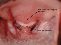 abnormalities
of the head. Procedures: Medical records and archived midsagittal
T2-weighted MRI images of brachycephalic and nonbrachycephalic dogs' heads
were reviewed. Group assignment was based on breed. Data collected included
weight, SPL and thickness, OBA, and the distance between the caudal
extremity of the soft palate and the basihyoid. Soft palate length and
thickness were adjusted on the basis of body weight. Results: Brachycephalic
dogs had significantly thicker soft palates and lower OBAs, compared with
findings for nonbrachycephalic dogs. There was a significant negative
correlation (r2 = 0.45) between OBA and soft palate thickness. The
correlation between SPL and OBA was less profound (r2 = 0.09). The distance
between the caudal extremity of the soft palate and the basihyoid was
shorter in brachycephalic dogs than in nonbrachycephalic dogs. The
percentage of epiglottis–soft palate overlap significantly decreased with
increasing OBA (r2 = 0.31). Conclusions & Clinical Relevance: Results
indicated that MRI images can be consistently used to assess anatomic
landmarks for measurement of SPL and thickness, OBA, and soft
palate-to-epiglottis distance in brachycephalic and nonbrachycephalic dogs.
The percentage of epiglottis–soft palate overlap was significantly greater
in brachycephalic dogs and was correlated to the degree of brachycephalia.
abnormalities
of the head. Procedures: Medical records and archived midsagittal
T2-weighted MRI images of brachycephalic and nonbrachycephalic dogs' heads
were reviewed. Group assignment was based on breed. Data collected included
weight, SPL and thickness, OBA, and the distance between the caudal
extremity of the soft palate and the basihyoid. Soft palate length and
thickness were adjusted on the basis of body weight. Results: Brachycephalic
dogs had significantly thicker soft palates and lower OBAs, compared with
findings for nonbrachycephalic dogs. There was a significant negative
correlation (r2 = 0.45) between OBA and soft palate thickness. The
correlation between SPL and OBA was less profound (r2 = 0.09). The distance
between the caudal extremity of the soft palate and the basihyoid was
shorter in brachycephalic dogs than in nonbrachycephalic dogs. The
percentage of epiglottis–soft palate overlap significantly decreased with
increasing OBA (r2 = 0.31). Conclusions & Clinical Relevance: Results
indicated that MRI images can be consistently used to assess anatomic
landmarks for measurement of SPL and thickness, OBA, and soft
palate-to-epiglottis distance in brachycephalic and nonbrachycephalic dogs.
The percentage of epiglottis–soft palate overlap was significantly greater
in brachycephalic dogs and was correlated to the degree of brachycephalia.
The prevalence of dynamic pharyngeal collapse is high in brachycephalic dogs undergoing videofluoroscopy. Rachel E. Pollard, Lynelle R. Johnson, Stanley L.Marks. Vet. Radial Ultrasound. April 2018; doi: 10.1111/vru.12655. Quote: The aim of this retrospective study was to determine the frequency of pharyngeal collapse ina large group of brachycephalic dogs undergoing videofluoroscopic assessment of swallowingor airway diameter. We hypothesized that brachycephalic dogs would have pharyngeal collapsemore frequently than dolichocephalic or mesocephalic dogs with or without airway collapse. Themedical records database was searched for brachycephalic dogs undergoing videofluoroscopyof swallowing or airway diameter between January 1, 2006 and December 31, 2015. A cohortof dolichocephalic/mesocephalic dogs with videofluoroscopically confirmed airway collapse wasage and time matched for comparison. A control group of dolichocephalic/mesocephalic dogsthat did not have documented airway collapse was also evaluated. All fluoroscopic studies wereassessed by a board certified veterinary radiologist for the presence and degree of pharyngealcollapse. Results demonstrate that pharyngeal collapsewas significantly more common in brachycephalicdogs (58/82; 72%) than in nonbrachycephalic dogs with (7/25; 28%) and without (2/30;7%) airway collapse. Pharyngeal collapse is more prevalent in brachycephalic dogs undergoingvideofluoroscopy than in dolichocephalic/mesocephalic dogs with or without airway collapse.
Sleep disordered breathing in the Cavalier King Charles Spaniel: a case series. Tom Hinchliffe, Nai-Chieh Liu, Jane Ladlow. BSAVA Congress 2018 Proceedings. April 2018;pp. 479-480. Quote: Objectives: Obstructive sleep apnoea has been widely investigated in human medicine, but relatively few studies have described this condition in veterinary species; to date it has only been reported in English Bulldogs, French Bulldogs and Pugs. The aim of this study was to report the clinical features, diagnosis, management and outcome of five cases of obstructive sleep apnoea in the Cavalier King Charles Spaniel (CKCS). Methods: A retrospective case series was created using hospital records over five years, followed up via recheck appointments and a telephone questionnaire. The inclusion criteria consisted of CKCSs presenting primarily for sleep disorders, which were treated surgically for obstructive anatomical pathology. Results: All five cases presented with stertorous breathing, choking and apnoea during sleep. All cases had a number of co-morbidities: 5/5 had eosinophilic stomatitis and otitis media, 4/5 had mitral valve insufficiency and 3/5 had syringomyelia. CT and rhinoscopy revealed that 5/5 had aberrant nasal turbinates, nasal septal deviation and soft palate thickening and 3/5 had nasopharyngeal thickening and tracheal collapse. Treatment consisted of a combination of laser turbinectomy, folding flap palatoplasty, tonsillectomy, laryngeal sacculectomy and cuneiform process resection. All cases had an improvement in both the incidence and severity of sleep apnoea within a week, with 4/5 cases achieving complete resolution. Conclusions: This study concludes that obstructive sleep apnoea is a condition that may affect the CKCS and all five cases described here improved following surgical intervention. Consequntly, sleep disordered breathing is a presentation that veterinary surgeons should be aware of in this breed. (See this May 2019 article on the same study.)
Long-term outcome of permanent tracheostomy in 15 dogs with severe laryngeal collapse secondary to brachycephalic airway obstructive syndrome. Matteo Gobbetti, Stefano Romussi, Paolo Buracco, Valerio Bronzo, Samuele Gatti, Matteo Cantatore. Vet. Surg. June 2018;doi:10.1111/vsu.12903. Quote: Objective: To report the long‐term outcome of permanent tracheostomy for the management of severe laryngeal collapse secondary to brachycephalic airway obstructive syndrome. Study design: Retrospective case series. Animals: Fifteen brachycephalic dogs with severe laryngeal collapse [including one cavalier King Charles spaniel] treated with permanent tracheostomy. Methods: Follow-up data were obtained from medical records or via telephone conversation with the owners. The Kaplan-Meier estimator was used to calculate median survival time. Death was classified as related or unrelated to tracheostomy surgery. Complications were classified as major when they were life-threatening or required revision surgery. Owners were asked to classify the postoperative quality of life as improved, unchanged, or worse and the management of the stoma as simple or demanding. Results: The median survival time was 100 days [the CKCS survied 143 days]. Major complications were diagnosed in 12 of 15 (80%) dogs, resulting in death in 8 (median survival time 15 days) and revision surgery in 4 dogs [including the CKCS]. Seven of 15 (47%) dogs died of unrelated causes [including the CKCS] or were alive at the end of the study (median survival time 1982 days). The postoperative quality of life of 9 dogs was judged as markedly improved [including the CKCS]. Stoma management was defined as simple in 8 dogs and demanding in 4. Conclusion: Permanent tracheostomy was associated with a high risk of complications and postoperative death in brachycephalic dogs. However, long-term survival (exceeding 5 years) with a good quality of life was documented in 5 of 15 dogs [including the CKCS]. Clinical significance: Permanent tracheostomy is a suitable salvage option in brachycephalic dogs with severe laryngeal collapse that did not improve following more conservative surgeries.
Risk of anesthesia-related complications in brachycephalic dogs. Michaela Gruenheid, Turi K. Aarnes, Mary A. McLoughlin, Elaine M. Simpson, Dimitria A. Mathys, Dixie F. Mollenkopf, Thomas E. Wittum. JAVMA. August 2018;253(3):301-306. Quote: Objective: To determine whether brachycephalic dogs were at greater risk of anesthesia-related complications than nonbrachycephalic dogs and identify other risk factors for such complications. Design: Retrospective cohort study. Animals: 223 client-owned brachycephalic dogs [none were cavalier King Charles spaniels] undergoing general anesthesia for routine surgery or diagnostic imaging during 2012 and 223 nonbrachycephalic client-owned dogs matched by surgical procedure and other characteristics. Procedures: Data were obtained from the medical records regarding dog signalment, clinical signs, anesthetic variables, surgery characteristics, and complications noted during or following anesthesia (prior to discharge from the hospital). Risk of complications was compared between brachycephalic and nonbrachycephalic dogs, controlling for other factors. Results: Perianesthetic (intra-anesthetic and postanesthetic) complications were recorded for 49.1% (n = 219) of all 446 dogs (49.8% [111/223] of brachycephalic and 48.4% [108/223] of nonbrachycephalic dogs), and postanesthetic complications were recorded for 8.7% (39/446; 13.9% [31/223] of brachycephalic and 3.6% [8/223] of nonbrachycephalic dogs). Factors associated with a higher perianesthetic complication rate included brachycephalic status and longer (vs shorter) duration of anesthesia; the risk of perianesthetic complications decreased with increasing body weight and with orthopedic or radiologic procedures (vs soft tissue procedures). Factors associated with a higher postanesthetic complication rate included brachycephalic status, increasing American Society of Anesthesiologists status, use of ketamine plus a benzodiazepine (vs propofol with or without lidocaine) for anesthetic induction, and invasive (vs noninvasive) procedures. Conclusions & Clinical Relevance: Controlling for other factors, brachycephalic dogs undergoing routine surgery or imaging were at higher risk of peri- and postanesthetic complications than nonbrachycephalic dogs. Careful monitoring is recommended for brachycephalic dogs in the perianesthetic period.
RETURN TO TOP
2019
Consequences and Management of Canine Brachycephaly in Veterinary Practice: Perspectives from Australian Veterinarians and Veterinary Specialists. Anne Fawcett, Vanessa Barrs, Magdoline Awad, Georgina Child, Laurencie Brunel, Erin Mooney, Fernando Martinez-Taboada, Beth McDonald, Paul McGreevy. Animals. January 2019;9(1):3. Quote: This article, written by veterinarians whose caseloads include brachycephalic dogs, argues that there is now widespread evidence documenting a link between extreme brachycephalic phenotypes and chronic disease, which compromises canine welfare. This paper is divided into nine sections exploring the breadth of the impact of brachycephaly on the incidence of disease, as indicated by pet insurance claims data from an Australian pet insurance provider, the stabilization of respiratory distress associated with brachycephalic obstructive airway syndrome (BOAS), challenges associated with sedation and the anaesthesia of patients with BOAS; effects of brachycephaly on the brain and associated neurological conditions, dermatological conditions associated with brachycephalic breeds, and other conditions, including ophthalmic and orthopedic conditions, and behavioural consequences of brachycephaly. In the light of this information, we discuss the ethical challenges that are associated with brachycephalic breeds, and the role of the veterinarian. ... Petsure Australia has collected relevant prevalent and actuarial data over the years, in the process of administering its core business of providing pet insurance policies. This includes the following brachycephalic breeds from their database in its ongoing in-house study: Affenpinscher, American Bulldog, Australian Bulldog, Australian Bulldog Miniature, Boston Terrier, Boxer, British Bulldog, Cavalier King Charles Spaniel (CKCS), Dogue De Bordeaux, French Bulldog, Griffon, Griffon Brabancon, Griffon Bruxellois, Lhasa Apso, Mastiff, Neopolitan Mastiff, Pekingese, Pug and Shih Tzu. ... In addition, individual breeds have specific dermatological disorders, including primary secretory otitis media (PSOM) in the Cavalier King Charles Spaniel. ... CM has been described most commonly in the CKCS, and is reported to occur in 100%of dogs of this breed. ... CKCSs and Griffons Bruxellois are highly predisposed to SM, with a prevalence in dogs without recognised clinical signs being estimated at 25% at 12 months of age, 70% at six years or older in CKCSs ... (of varying ages). The incidence of dogs showing clinical signs associated with CM and/or SM is much lower. CM and SM have a moderately high heritability in CKCSs, and genetic factors are likely in other affected breeds and many of their crosses. SM is almost certainly associated directly with skull conformation. The mode of inheritance is unclear, but it does not appear to be simple. Phenotypic screening of CKCSs and Griffons Bruxellois in the last 10 years seems to have reduced the incidence of severely affected dogs in breeding lines, where screening with magnetic resonance imaging (MRI) has been undertaken, but has not eliminated the condition. ... Two significant risk factors that are associated with the development of CM/SM in the conformation of the CKCS are the extent of the brachycephaly, and the distribution of the cranium (higher cephalic index with a broader, shorter head and rostrocaudal doming. ... Brachycephalic breeds may experience a higher incidence of orthopaedic conditions. ... The prevalence of patellar luxation was ... 3.8% in CKCS ..., compared with an overall prevalence of 1.3%. ... In summary, dogs with BOAS do not enjoy freedom from discomfort, nor freedom from pain, injury, and disease, and they do not enjoy the freedom to express normal behaviour. According to both deontological and utilitarian ethical frameworks, the breeding of dogs with BOAS cannot be justified, and further, cannot be recommended, and indeed, should be discouraged by veterinarians.
Sleep-disordered breathing in the Cavalier King Charles spaniel: A case series. Tom A. Hinchliffe, Nai-Chieh Liu, Jane Ladlow. Vet. Surg. May 2019;48(4):497-504. Quote: Objective: To describe sleep-disordered breathing (SDB) in the Cavalier King Charles spaniel (CKCS). Study design Retrospective case series. Animals: Five client-owned dogs referred for SDB. Methods: Medical records were reviewed including recheck appointments and routine preoperative and postoperative questionnaires. Whole-body barometric plethysmography was used to categorize SDB. Results: All dogs presented with multiple episodes of stertorous breathing, choking, and apnea during sleep. Severe nasal septal deviation, aberrant nasal turbinates, and soft palate elongation and thickening were noted on computed tomography and rhinoscopy of each dog. Whole-body barometric plethysmography measurements during sleep (in 3 dogs) documented periods of choking, snoring, and apnea. Treatment combined laser turbinectomy, folding flap palatoplasty, tonsillectomy, laryngeal sacculectomy, and cuneiform process resection. All dogs improved in terms of incidence and severity of sleep apnea within 1 week, with 4 of 5 dogs achieving complete resolution. Conclusion: The objective measurements used to characterize SDB in this population of CKCS provided some evidence to support an obstructive cause for this condition, which improved with surgical treatment. Clinical significance Sleep-disordered breathing in the CKCS is a different clinical presentation of brachycephalic obstructive airway syndrome. Our finding of intranasal abnormalities in these 5 dogs with SDB provides justification for future research into its clinical significance. (See this April 2018 abstract of the same study.)
Postoperative regurgitation in dogs after upper airway surgery to treat brachycephalic obstructive airway syndrome: 258 cases (2013-2017). Joy V. H. Fenner, Robert J. Quinn, Jackie L. Demetriou. Vet. Surgery. July 2019; doi: 10.1111/vsu.13297 Quote: Objective: To determine the incidence of and risk factors for regurgitation in dogs within 24 hours of surgical management of brachycephalic obstructive airway syndrome (BOAS). Study design: Retrospective single center study of dogs undergoing BOAS surgery over four years (2013-2017). Animals: Two hundred fifty-eight client-owned dogs referred for surgical intervention for BOAS. [20 cavalier King Charles spaniels -- 8%] Methods: Electronic medical records were searched for dogs that had undergone surgery for BOAS at a UK specialist referral hospital. Data were assessed by using univariable binomial logistic regression; confounding factors were then identified in a multivariable model. Results: There was an increase in the proportion of dogs that regurgitated while hospitalized preoperatively vs during the first 24 hours postoperatively, from 28 (10.9%) to 89 (34.5%), respectively (P<.0001). History of regurgitation (P =.017, odds ratio [OR] 2.539, 95% confidence interval [CI] 1.178-5.469) and age (P =.008, OR 0.712, 95% CI 0.553-0.916) were detected as risk factors for postoperative regurgitation. For every 1-year increase in age, the odds of experiencing postoperative regurgitation were reduced by 28.8%. [None of the CKCSs had any post-operative regurgitation.] Conclusion: Corrective surgery for BOAS was associated with a marked incidence of postoperative regurgitation. Younger dogs and those with a history of regurgitation were predisposed to postoperative regurgitation. Clinical significance: The increased frequency of regurgitation after surgical treatment of BOAS, especially in younger dogs, provides justification for counseling owners regarding this postoperative complication.
RETURN TO TOP
2020
Brachycephalic airway syndrome: management of post-operative respiratory complications in 248 dogs. B Lindsay, D Cook, J-M Wetzel, S Siess, P Moses. Austral. Vet. J. February 2020; doi: 10.1111/avj.12926. Quote: Objective: As ownership of brachycephalic dog breeds rises, the surgical correction of components of brachycephalic airway syndrome (BAS) is increasingly recommended by veterinarians. This study's objective was to describe the incidence of, and strategies for the management of post-operative respiratory complications in brachycephalic dogs undergoing surgical correction of one or more components of BAS. Methods: Medical records of 248 brachycephalic dogs [42 cavalier King Charles spaniels (CKCS)(16.9%)]treated surgically for BAS were retrospectively reviewed for demographic information, procedures performed, post-operative complications and treatment implemented, hospitalisation time, and necessity for further surgery. Results: Pugs, Cavalier King Charles Spaniels and British Bulldogs were the most commonly encountered breeds. Dogs which experienced a complication were significantly older (mean was 5.5 years, compared with 4.1 years [P < 0.01]). ... CKCSs were found to present significantly older than the rest of the population (mean age 5.8 years). ... Table 1: Number of dogs with various compoments of brachycephalic airway syndrome treated surgically: Elongated soft palate - 242 dogs (97.6%); hyperplastic tonsillar tissue - 106 dogs (42.7%); everted saccules - 103 dogs (41.5%); stenotic nares - 97 dogs (39.1%). ... Fifty-eight dogs (23.4%) had complications which included: dyspnoea managed with supplemental oxygen alone (7.3%, n = 18), dyspnoea requiring anaesthesia and re-intubation (8.9%, n = 22), dyspnoea necessitating treatment with a temporary tracheostomy (8.9%, n = 22), aspiration pneumonia (4%, n = 10), and respiratory or cardiac arrest (2.4%, n = 6). Five of the 22 dogs requiring anaesthesia and re-intubation deteriorated 12 or more hours after post-surgical anaesthetic recovery. The overall mortality rate in this study was 2.4% (n = 6). Age, concurrent airway pathology, and emergency presentation significantly predicted post-operative complications. Conclusion: Our data show the importance of close monitoring for a minimum of 24 h following surgery by an experienced veterinarian or veterinary technician. Surgical intervention for BAS symptomatic dogs should be considered at an earlier age as an elective procedure, to reduce the risk of post-operative complications.
“Forever young” — Postnatal growth inhibition of the turbinal skeleton in brachycephalic dog breeds (Canis lupus familiaris). Franziska Wagner, Irina Ruf. Anat. Rec. May 2020; doi: 10.1002/ar.24422 Quote: In short snouted (brachycephalic) dogs (Canis lupus familiaris ), several genetic mutations cause postnatal growth inhibition of the viscerocranium. Thus, for example, the pug keeps a snub nose like that observed in neonate dogs in general. However, little is known how far intranasal structures like the turbinal skeleton are also affected. In the present study, we provide the first detailed morphological and morphometric analyses on the turbinal skeleton of pug, Japanese chin, pekingese, King Charles spaniel, and Cavalier. ... The brachycephalic King spaniel and the moderate brachycephalic to mesaticephalic Cavalier serve as an example for the reverse breeding, namely the elongation of the face. ... By this, the following hypotheses are evaluated: ... (c) the fast secondary elongation of the snout in the Cavalier exhibits a similar pattern to the pug in reverse. Based on the ancestral stage predetermined by the King spaniel, within the enlarged nasal cavity the still simplified turbinals of the Cavalier have a smaller IAT and a looser arrangement, that is, a smaller SDEN [turbinal surface density]. ... In order to elucidate how a shortened snout affects turbinal shape, size, and density, our sample covers different degrees of brachycephaly. Macerated skulls of 1 juvenile and 17 adult individuals [including 2 cavalier King Charles spaniels -- NMBE1059207 & NMBE1062998] were investigated by μCT and virtual 3D reconstructions. In addition, histological serial sections of two prenatal and one neonate whippet were taken into account. All investigated postnatal stages show three frontoturbinals and three ethmoturbinals similar to longer snouted breeds, whereas the number of interturbinals is reduced. The shape of the entire turbinal skeleton simplifies with decreasing snout length, that is, within a minimized nasal cavity the turbinals decrease proportionally in surface area and surface density due to a looser arrangement. We interpret these apparent reductions as a result of spatial constraint which affects postnatal appositional bone growth and the position of the turbinals inside the nasal cavity. The turbinal skeleton of brachycephalic dogs arrests at an early ontogenetic stage, corresponding with previous studies on the dermal bones. Hence, we assume an association between the growth of intranasal structures and facial elongation. ... Our current study proves that the medium snouted ancient pug ZMB_MAM 30980 and the Cavalier specimen correspond to this size relation, whereas the more extreme brachycephalic Toy dogs are similar to the juvenile pattern. ... The influence of ontogeny in association with facial length growth on the development of the turbinal skeleton further explains the latter's simple morphology in the short snouted modern pug on the one hand, and the complex shape in the moderate brachycephalic to mesaticephalic Cavalier on the other hand. As expected, the ancient Toy breeds (chin, pekingese, King spaniel, ancient pug) have a smaller IAT and lower SDEN than is reflected by the grundplan . Contrasting our other two hypotheses, fast evolutionary changes, like the artificial shortening (modern pug) or elongation (Cavalier) of the face, have no or little influence on the development of the turbinal skeleton. ... Our study clearly demonstrates that the rapid change of facial shape in dogs first affects the number of the least developed turbinals, especially the additional interturbinals. ... The species-specific number of fronto-, ethmo-, and prominent interturbinals is forced to develop within the smaller space of the shortened snout in brachycephalic dogs. ...The genetic mutations, which restrict the facial length growth in brachycephalic breeds, also affect the development of the turbinals and determine both, the endochondral ossification of the fetal cartilaginous turbinals as well as the intramembraneous ossification of their accessory lamellae. The reduced space within the nasal cavity effects directly the arrangement of the turbinal skeleton and forces certain parts to avoid the restricted space as aberrant turbinals.
Sleep in the dog: comparative, behavioral and translational relevance. Róbert Bódizs1, Anna Kis, Márta Gácsi, József Topál. Current Opin. Behavior Sci. June 2020; doi: 10.1016/j.cobeha.2019.12.006. Quote: The dog (Canis familiaris) is a promising non-invasive translational model of human cognitive neuroscience including sleep research. Studies on the relationship between sleep and cognition in dogs and other canines are only just emerging, but still very scarce. Here we provide insight into canine sleep and sleep-related physiological and cognitive/behavioral phenomena. We show that dogs do not only fulfil all behavioral and polygraphic criteria of sleep, but are characterized by sleep homeostasis, diurnal pattern of activity, circadian rhythms, ultradian sleep cycles, socio-ecologically and environmentally shaped wake-sleep structure, sleep-related memory improvement, as well as specific sleep disorders. Developmental patterns of sleep-related physiological indices, as well as parallel trends in age-dependent changes in cognition and sleep were evidenced in dogs. • All behavioral and polygraphic criteria of (NREM and REM) sleep are met in the dog. • Dogs are diurnal: weak circadian modulation and assumed adaptation to human rhythms. • Pre-sleep experiences, location and socio-ecological factors affect canine sleep. • Sleep may contribute to dogs’ memory consolidation. • Development and aging affect sleep and cognition in dogs. ... Sleep disordered breathing is associated with episodes of O2 desaturation and loud snoring during sleep, as well as daytime hypersomnolence, sluggishness, and shortened sleep latency. The English bulldog, the Cavalier King Charles spaniel, as well as other brachycephalic breeds are most commonly affected. The English bulldog has been proposed as a natural model of sleep-disordered breathing.
Dorsal offset rhinoplasty for treatment of stenotic nares in 34 brachycephalic dogs. Vanna M. Dickerson, Corinne M. B. Dillard, Janet A. Grimes, Mandy L. Wallace, Jonathan F. McAnulty, Chad W. Schmiedt. Vet. Surg. August 2020; doi: 10.1111/vsu.13504. Quote: Objective: To describe the technique, outcome, and owner satisfaction associated with dorsal offset rhinoplasty (DOR) to treat stenotic nares in brachycephalic dogs. Study design: Retrospective case series. Animals: Thirty-four client-owned dogs. ... The most common breed was the English bulldog (n = 18), followed by the French bulldog (n = 6), Boston terrier (n = 4), pug (n = 2), shih tzu (n = 2), Japanese chin (n = 1), and Cavalier King Charles spaniel (n = 1). ... Methods: Medical records of dogs treated with DOR at a veterinary teaching hospital over a 6-year period were identified. Dorsal offset rhinoplasty was defined as removal of a dorsal wedge of nasal planum from each naris with apposition of the rostral abaxial tissue to the caudal axial tissue, resulting in translocation of the alar cartilage in both median and dorsal planes. Immediate and postoperative complications were recorded. Owners were asked to report any complications with healing of the nares and to score their satisfaction with the appearance of the nares. Results: Thirty-four dogs met the inclusion criteria. Twenty-nine (85%) dogs were examined a median of 402.5 days (range, 23-2042) postoperatively, with no major complications related to the rhinoplasty recorded. Eighteen owners responded a median of 701 days (range, 37-1622) postoperatively. One owner reported that self-trauma led to collapse of one naris. One owner reported collapse of both nares within 4 years; timing and cause were unknown. Sixteen of 17 responding owners reported that they were very satisfied with the outcome of the rhinoplasty. The owner of the dog with the collapsed naris was very unsatisfied. One owner did not provide a satisfaction score. Conclusion: Owners were generally highly satisfied with DOR, and complications were uncommon. Clinical significance: This report describes an alternate technique to treat stenotic nares.
Unravelling the health status of brachycephalic dogs in the UK using multivariable analysis. D. G. O’Neill, C. Pegram, P. Crocker, D. C. Brodbelt, D. B. Church, R. M. A. Packer. Sci. Rep. October 2020; doi: 10.1038/s41598-020-73088-y. Quote: Brachycephalic dog breeds are regularly asserted as being less healthy than non-brachycephalic breeds. Using primary-care veterinary clinical data, this study aimed to identify predispositions and protections in brachycephalic dogs and explore differing inferences between univariable and multivariable results. All disorders during 2016 were extracted from a random sample of 22,333 dogs within the VetCompass Programme from a sampling frame of 955,554 dogs under UK veterinary care in 2016. ... There were 4,169 (18.74%) brachycephalic and 18,079 (81.26%) non-brachycephalic types. ... The brachycephalic group included 34 individual breeds ... The most common brachycephalic breeds were Chihuahua (n = 955, 22.91%), Shih-tzu (795, 19.07%) and Cavalier King Charles Spaniel (435, 10.43%). ... The degree (or severity) of brachycephaly varies between breeds (a bulldog may be considered as more severely brachycephalic than a Cavalier King Charles Spaniel) but there can also be considerable variation in brachycephaly within breeds. Shifting the median severity of brachycephaly towards a longer skull shape within breeds has been suggested as one option to reduce the prevalence of disorders directly linked to brachycephaly while still retaining these breeds within the overall dog population. ... Univariable and multivariable binary logistic regression modelling explored brachycephaly as a risk factor for each of a series of common disorders. Brachycephalic dogs were younger, lighter and less likely to be neutered than mesocephalic, dolichocephalic and crossbred dogs. Brachycephalic differed to non-brachycephalic types in their odds for 10/30 (33.33%) common disorders. Of these, brachycephalic types were predisposed for eight disorders and were protected for two disorders. ...The most common precise disorders (i.e. greatest prevalence) in the brachycephalic types were periodontal disease (n = 485, prevalence = 11.63%), otitis externa (303, 7.27%), obesity (266, 6.38%), anal sac impaction (249, 5.97%), overgrown nail(s) (212, 5.09%), diarrhoea (143, 3.43%) and heart murmur (3.43%). There were eight precise disorders with the higher odds in multivariable logistic regression analysis (i.e. disorder predisposition) for brachycephalic types compared with non-brachycephalic types: corneal ulceration, heart murmur, umbilical hernia, pododermatitis, skin cyst, patellar luxation, otitis externa and anal sac impaction. Two precise disorders had reduced odds for brachycephalic types in multivariable analysis: undesirable behaviour and claw injury. ... This study provides strong evidence that brachycephalic breeds are generally less healthy than their non-brachycephalic counterparts. Results from studies that report only univariable methods should be treated with extreme caution due to potential confounding effects that have not been accounted for during univariable study design or analysis. ... Conclusion: This study provides strong evidence to support the common assertion that brachycephalic breeds are generally less healthy than their non-brachycephalic counterparts in relation to total disorder counts and specific common conditions recorded. Potential solutions to some of these health problems are likely to require conformational change to current skull shapes averages for many breeds; however, many other health problems will require targeted action at the individual breed level, owing to large differences in individual breed predispositions to disorders. Results from studies that report only univariable methods should be treated with extreme caution due to potential confounding effects that have not been accounted for during study design or analysis.
“Forever young” — Postnatal growth inhibition of the turbinal skeleton in brachycephalic dog breeds (Canis lupus familiaris). Franziska Wagner, Irina Ruf. Anat. Rec. December 2020; doi: 10.1002/ar.24422. Quote: In short snouted (brachycephalic) dogs (Canis lupus familiaris), several genetic mutations cause postnatal growth inhibition of the viscerocranium [facial bones]. Thus, for example, the pug keeps a snub nose like that observed in neonate dogs in general. However, little is known how far intranasal structures like the turbinal [nasal] skeleton are also affected. ... The brachycephalic King spaniel and the moderate brachycephalic to mesaticephalic Cavalier serve as an example for the reverse breeding, namely the elongation of the face. ... In the present study, we provide the first detailed morphological and morphometric analyses on the turbinal skeleton of pug, Japanese chin, pekingese, King Charles spaniel, and Cavalier. In order to elucidate how a shortened snout affects turbinal shape, size, and density, our sample covers different degrees of brachycephaly. Macerated skulls of 1 juvenile and 17 adult individuals [including two cavalier King Charles spaniels] were investigated by μCT and virtual 3D reconstructions. In addition, histological serial sections of two prenatal and one neonate whippet were taken into account. All investigated postnatal stages show three frontoturbinals and three ethmoturbinals similar to longer snouted breeds, whereas the number of interturbinals is reduced. The shape of the entire turbinal skeleton simplifies with decreasing snout length, that is, within a minimized nasal cavity the turbinals decrease proportionally in surface area and surface density due to a looser arrangement. We interpret these apparent reductions as a result of spatial constraint which affects postnatal appositional bone growth and the position of the turbinals inside the nasal cavity. The turbinal skeleton of brachycephalic dogs arrests at an early ontogenetic stage, corresponding with previous studies on the dermal bones. Hence, we assume an association between the growth of intranasal structures and facial elongation. ... Contrasting our other two hypotheses, fast evolutionary changes, like the artificial shortening (modern pug) or elongation (Cavalier) of the face, have no or little influence on the development of the turbinal skeleton.
RETURN TO TOP
2021
Sleep disorders in dogs: A pathophysiological and clinical review. Alejandra Mondino, Luis Delucchi, Adam Moeser, Sofía Cerdá-González, Giancarlo Vanini. Topics in Companion Anim. Med. February 2021; doi: 10.1016/j.tcam.2021.100516. Quote: Sleep is a fundamental process in mammals, including domestic dogs. Disturbances in sleep affect physiological functions like cognitive and physical performance, immune response, pain sensation and increase the risk of diseases. In dogs, sleep can be affected by several conditions, with narcolepsy, REM sleep behavior disorder and sleep breathing disorders being the most frequent causes. Furthermore, sleep disturbances can be a symptom of other primary diseases where they can contribute to the worsening of clinical signs. This review describes reciprocally interacting sleep and wakefulness promoting systems and how their dysfunction can explain the pathophysiological mechanisms of sleep disorders. ... Cavalier King Charles Spaniel (CKCS) ... can be affected with SBD [sleep-related breathing disorders]. The severity of the disorder can range from partial obstructive snoring to apnea. ... CKCS, one of the breeds affected by SDB, also have a high prevalence of Chiari-like malformations (Caudal Occipital Malformation Syndrome, (COMS). In fact, 60% of the CKCS with SDB studied by Hinchliffe et al., (2018) had syringomyelia, which can occur secondary to COMS among other craniocervical junction disorders. However, these dogs all responded well to surgical interventions of the upper airway, suggesting that the SDB was at least partially explained by OSA [obstructive sleep apnea]. In dogs with OSA the anatomic features responsible for the disorder should be determined by complete oropharyngeal examination, endoscopy, and computed tomography as needed. ... Additionally, this work discusses the clinical characteristics, diagnostic tools and available treatments for these disorders while highlighting areas in where further studies are needed so as to improve their treatment and prevention.
The Need for Head Space: Brachycephaly and Cerebrospinal Fluid Disorders. Clare Rusbridge, Penny Knowler. Life. February 2021; doi: 10.3390/life11020139. Quote: Brachycephalic dogs remain popular, despite the knowledge that this head conformation is associated with health problems, including airway compromise, ocular disorders, neurological disease, and other co-morbidities. There is increasing evidence that brachycephaly disrupts cerebrospinal fluid [CSF] movement and absorption, predisposing ventriculomegaly, hydrocephalus, quadrigeminal cistern expansion, Chiari-like malformation, and syringomyelia. In this review, we focus on cerebrospinal fluid physiology and how this is impacted by brachycephaly, airorhynchy, and associated craniosynostosis [premature closure of cranial sutures, preventing continued growth to accommodate the growing size of the brain]. ... As the majority of CSF is absorbed through lymphatics in the skull base and via the olfactory bulbs, the balance between CSF production and absorption may be impacted by craniofacial hypoplasia and cranial base shortening. ... Respiration drives CSF movement through the ventricular system. ... [O]ne of the most important skull base foramen [openings] is the jugular, situated between the temporal and occipital bone. The jugular foramen provides passage to cranial nerves IX, X, XI, sigmoid sinus, and lymphatics. Cavalier King Charles spaniels with syringomyelia associated with Chiari-like malformation have smaller volume jugular foramina compared to Cavalier King Charles spaniels without syringomyelia. However, direct causality between smaller jugular foramen and syringomyelia has not been proven. ... Cavalier King Charles spaniels with syringomyelia associated with Chiari malformation have reduced volume caudal cranial fossa dorsal sinuses. ... Dogs and cats with neonatal characteristics of a reduced muzzle and brachycephaly have impaired CSF circulation, which predisposes ventriculomegaly, hydrocephalus, quadrigeminal cistern expansion, Chiari-like malformation associated pain, and syringomyelia. Reduction of the lymphatic absorption of CSF through the nasal and skull base lymphatics, in conjunction with restriction of CSF movement through the lateral apertures and craniocervical junction, are hypothesized to be key features. Cranial venous stenosis, spinal canal conformation, chronic intermittent hypoxia, and thoracic cavity pressure gradients may contribute. The ethics of choosing to breed animals with this predisposition is questionable, despite their popularity as companion animals. Education of the pet-buying public, avoiding breeding extreme animals, and screening at-risk breeding stock is recommended.
Management of sleep disordered breathing in a dog using continuous positive airway pressure. Waylon Wiseman, A. Rosenblatt, M. Kunga. Aust. Vet. Pract. April 2021;51(1):19-25. Quote: A 4-year-old Cavalier King Charles spaniel presented for evaluation of disruptive apnoeic episodes, experienced every 10-15 minutes during sleep. Surgical intervention included bilateral tonsillectomy, bilateral laryngeal sacculectomy, and unilateral cricoarytenoid lateralisation. Minimal improvement in the frequency and severity of episodes of sleep disordered breathing were noted. ... We hypothesise that our patientcontinued to suffer SDB, despite corrective airway surgery with good clinical response while awake, due to DPC when sleeping, secondary to muscle hypotonia as seen in humans. ... The patient was subsequently fitted with a continuous positive airway pressure [CPAP] device at night and responded well to this intervention, allowing the dog to sleep through the night. Four years after the initial presentation, the patient underwent fluoroscopic large airway assessment and was diagnosed with dynamic pharyngeal collapse [DPC]. ... We then retrospectively confirmed partial pharyngeal collapse using videofluoroscopy. ... To the authors’ knowledge, this is the first report of the use of continuous positive airway pressure in the management of sleep disordered breathing in dogs and represents a novel management strategy for dogs suffering from this condition that respond poorly to airway surgery.
RETURN TO TOP
2022
Nasal Septum Deviation in dog breeds predisposed to Chiari-like malformation associated pain and syringomyelia. Clare Rusbridge, J. R. Rusbridge. BSAVA Congress 2022. March 2022. Quote: Nasal Septum deviation in breeds predisposed to Chiari-like malformation associated pain and syringomyelia. Objectives: Nasal septum deviation (NSD) and Chiari-like malformation pain and syringomyelia (CMSM) are common in flat-faced brachycephalic dogs. NSD may be clinically irrelevant but has been associated with sleep disordered breathing (SDB) in humans and dogs. SDB affects glymphatic drainage of cerebrospinal fluid which could predispose CMSM. This study investigated possible correlation between NSD and CMSM Methods Study population - 76 dogs with head CT [computed tomography] and known CMSM status (based on MRI and medical history). Comprising 53 CKCS [cavalier King Charles spaniels] (70%) (7 CM-N [CM-clinically normal] i.e. MRI determined breed-associated CM but clinically normal; 14 with CM-P [CM-associated pain] but no SM; 32 with SM); 12 Chihuahua (5 MRI and clinically normal, 7 with SM); 11 other brachycephalic toy dog breeds and crosses (10 with SM). Transverse CT of the nasal cavity in bone windows was reviewed. Presence of a NSD was recorded. Previously described method of determining the angle of NSD was not reproducible and consequently abandoned. The NSD ratio calculated (maximum distance of NSD from midline/ distance from midline to nasal cavity wall). Results NSD was almost ubiquitous; Only 3 dogs had a midline nasal septum without deviation (1 Chihuahua, 1 CKCS, 1 Cavapoo; all 3 were CMSM affected). There was no correlation between NSD ratio and CMSM (signs of pain or presence of syringomyelia). Impact NSD is common in dog breeds predisposed to CMSM. Care should be taken before attributing clinical relevance. Association of NSD to CMSM is unproven. More extreme brachycephaly is associated with both NSD, CM-P and SM and therefore the conditions may co-exist. Airway obstruction, SDB and apnoea may be relevant in the pathogenesis of CMSM or be a important comorbidity. More sophisticated methods of investigation are recommended for example polysomnography to monitor respiratory effort.
RETURN TO TOP
2023
Echocardiographic parameters in French Bulldogs, Pugs and Boston Terriers with brachycephalic obstructive airways syndrome. M. Brložnik, A. Nemec Svete, V. Erjavec & A. Domanjko Petric. BMC Vet. Res. February 2023; doi: 10.1186/s12917-023-03600-9. Quote: Background: In this prospective study, we hypothesized that dogs with signs of brachycephalic obstructive airway syndrome (BOAS) would show differences in left and right heart echocardiographic parameters compared with brachycephalic dogs without signs of BOAS and non-brachycephalic dogs. Result: We included 57 brachycephalic (30 French Bulldogs, 15 Pugs, and 12 Boston Terriers) and 10 non-brachycephalic control dogs. Brachycephalic dogs had significantly higher ratios of the left atrium to aorta and mitral early wave velocity to early diastolic septal annular velocity; smaller left ventricular (LV) diastolic internal diameter index; and lower tricuspid annular plane systolic excursion index, late diastolic annular velocity of the LV free wall, peak systolic septal annular velocity, late diastolic septal annular velocitiy, and right ventricular global strain than non-brachycephalic dogs. ... French Bulldogs: LA/Ao: 1.56 (1.44-1.60); Pugs: LA/Ao: 1.49 (1.45-1.62); Boston Terriers: LA/Ao: 1.55 (1.50-1.68). ... French Bulldogs with signs of BOAS had a smaller diameter of the left atrium index and right ventricular systolic area index; higher caudal vena cava at inspiration index; and lower caudal vena cava collapsibility index, late diastolic annular velocity of the LV free wall, and peak systolic annular velocity of the interventricular septum than non-brachycephalic dogs. Conclusions: ... The results of this study showed significant differences in echocardiographic parameters between the dogs of the three brachycephalic breeds and non-brachycephalic dogs. In addition, there were significant differences in echocardiographic parameters between dogs with and without signs of BOAS. ... We found significant differences in echocardiographic parameters between dogs of the three brachycephalic breeds and non-brachycephalic dogs, implying that breed-specific echocardiographic reference values should be used in clinical practice. In addition, significant differences were observed between brachycephalic dogs with and without signs of BOAS. The observed echocardiographic differences suggest higher right heart diastolic pressures affecting right heart function in brachycephalic dogs with and without signs of BOAS, and several of the differences are consistent with findings in OSA patients. Most of the changes of the heart morphology and function can be attributed to brachycephaly alone and not to the symptomatic stage.
Description of a novel method for detection of sleep-disordered breathing in brachycephalic dogs. Iida Niinikoski, Sari-Leena Himanen, Mirja Tenhunen, Liisa Lilja-Maula, Minna M. Rajamäki. J. Vet. Intern. Med. May 2023; doi: 10.1111/jvim.16783. Quote: Background: Sleep-disordered breathing (SDB), defined as any difficulty in breathing during sleep, occurs in brachycephalic dogs. Diagnostic methods for SDB in dogs require extensive equipment and laboratory assessment. Objectives: To evaluate the usability of a portable neckband system for detection of SDB in dogs. We hypothesized that the neckband is a feasible method for evaluation of SDB and that brachycephaly predisposes to SDB. Animals: Twenty-four prospectively recruited client-owned dogs: 12 brachycephalic dogs [including 9 French bulldogs and 1 cavalier King Charles spaniel] and 12 control dogs of mesocephalic or dolicocephalic breeds. Methods: Prospective observational cross-sectional study with convenience sampling. Recording was done over 1 night at each dog's home. The primary outcome measure was the obstructive Respiratory Event Index (OREI), which summarized the rate of obstructive SDB events per hour. Additionally, usability, duration of recording, and snore percentage were documented. Results: Brachycephalic dogs had a significantly higher OREI value (Hodges-Lehmann estimator for median difference = 3.5, 95% confidence interval [CI] 2.2-6.8) and snore percentage (Hodges-Lehmann estimator = 34.2, 95% CI 13.6-60.8) than controls. A strong positive correlation between OREI and snore percentage was detected in all dogs (rs = .79). ... The results would potentially be different if the BD [brachycephalic dog] group consisted of dogs with longer snouts such as the Bullmastiff and the Cavalier King Charles Spaniel. However, in addition to the laryngeal area, also obstruction of the nasal cavity can result in SDB, as documented in the Cavalier King Charles Spaniel. ... The neckband system was easy to use. Conclusions and Clinical Importance: Brachycephaly is associated with SDB. The neckband system is a feasible way of characterizing SDB in dogs.
The epidemiology of upper respiratory tract disorders in a population of insured Swedish dogs (2011–2014), and its association to brachycephaly. M. Dimopoulou, K. Engdah, J. Ladlow, G. Andersson, Å. Hedhammar, E. Skiöldebrand, I. Ljungvall. Nature Sci. Repts. May 2023; doi: 10.1038/s41598-023-35466-0. Quote: Upper respiratory tract (URT) disorders are common in dogs but neither general nor breed-related epidemiological data are widely reported. This study´s aims were to describe the epidemiology of URT disorders in a Swedish population of dogs and to investigate whether brachycephalic breeds were overrepresented among high-risk breeds. A cohort of dogs insured by Agria Djurförsäkring in Sweden (2011–2014) was used to calculate overall and breed-specific incidence rate (IR), age at first URT diagnosis and relative risk (RR) for URT disorders. For breeds with high RR for URT disorders, co-morbidities throughout the dog’s insurance period and age at death were investigated. The cohort included approximately 450,000 dogs. URT disorders had an overall IR of 50.56 (95% CI; 49.14–52.01) per 10,000 dog years at risk. Among 327 breeds, the English bulldog, Japanese chin, Pomeranian, Norwich terrier and pug had highest RR of URT disorders. ... The boxer, CKCS [cavalier King Charles spaniel], Chihuahua, French bulldog, pug and standard poodle had high risk for co-morbidity both prior and after the first URT claim. ... The Boston terrier, boxer, CKCS, English and French bulldog and pug are brachycephalic breeds and affected to a variable degree by BOAS [brachycephalic obstructive airway syndrome]. ... The Chihuahua, CKCS, Pomeranian and Yorkshire terrier are breeds frequently affected with the URT disorder tracheal collapse. Although the brachycephalic breeds most often cited in association with URT disorders are the English and French bulldog, the pug and the Boston terrier, our study found that the Japanese chin, Pomeranian, CKCS and boxer also have an increased risk for URT disorders. Among 13 breeds with high RR for URT disorders in this study, eight (Boston terrier, boxer, CKCS, English bulldog, French bulldog, Japanese chin, Pomeranian, pug) are recognised as brachycephalic by most researchers. ... The brachycephalic conformation has been linked to an increased risk for URT disorders, and specifically BOAS, in the French and English bulldog, the CKCS, Boston terrier and the pug. ... Eight of 13 breeds with high RR for URT disorders were brachycephalic. The median age at first URT diagnosis was 6.00 years (interquartile range 2.59–9.78). French bulldogs with URT diagnoses had a significantly shorter life span (median = 3.61 years) than other breeds with URT diagnosis (median = 7.81 years). Dogs with high risk for URT disorders had more co-morbidities than average.
RETURN TO TOP
2024
Evaluation of risk factors for sleep-disordered breathing in dogs. Iida Niinikoski, Sari-Leena Himanen, Mirja Tenhunen, Mimma Aromaa, Liisa Lilja-Maula, Minna M. Rajamäki. J. Vet. Intern. Med. February 2024; doi: 10.1111/jvim.17019. Quote: Background: Brachycephalic dogs display sleep-disordered breathing (SDB). The risk factors for SDB remain unknown. Objectives: To identify risk factors for SDB. We hypothesized that brachycephaly, increasing severity of brachycephalic obstructive airway syndrome (BOAS), excess weight, and aging predispose to SDB. Animals: Sixty-three privately owned pet dogs were prospectively recruited: 28 brachycephalic [including 8 cavalier King Charles spaniels (28.5%)] and 35 normocephalic (mesaticephalic or dolicocephalic) dogs. ... We had an overrepresentation of Cavalier King Charles Spaniels, French Bulldogs and Labrador Retrievers. Brachycephalic dogs were emphasized as SDB has been reported in these. ... Methods: Prospective observational cross-sectional study with convenience sampling. Recording with the neckband was done over 1 night at each dog's home. The primary outcome measure was the obstructive respiratory event index (OREI). Body condition score (BCS) was assessed, and BOAS severity was graded for brachycephalic dogs. Results: Brachycephaly was a significant risk factor for high OREI value (ratio of the geometric means 5.6, 95% confidence interval [CI] 3.2-9.9; P<.001) but aging was not (1.1, 95% CI 1.0-1.2; P=.2). Excess weight, defined as a BCS of over 5/9, (3.5, 95% CI 1.8-6.7; P<.001) was a significant risk factor. In brachycephalic dogs, BOAS-positive class (moderate or severe BOAS signs) was a significant risk factor (2.5, 95% CI 1.1-5.6; P=.03). ... Interestingly, Chiari malformation, a structural defect of the skull resulting in herniation of the brain into the spinal canal, has been associated with CSA in humans.52 Chiari-like malformation is prominent in many brachycephalic breeds, including the Cavalier King Charles Spaniel, where the defect is ubiquitous, but also in Pugs. However, central apnea or hypopnea events, excluding physiologic events related to sighs, did not occur in our study group. In a previous study, the resolution of apneas after extensive surgical procedures also supports obstructive, not central, apnea events in the Cavalier King Charles Spaniels studied. ... Although BOAS severity grading has not been evaluated in brachycephalic breeds excluding the extremely brachycephalic English Bulldog, French Bulldog and Pug, we performed it similarly in the other brachycephalic breeds. Only 1 of the 9 BOAS positive dogs represented a breed other than the 3 aforementioned breeds. This individual was a Cavalier King Charles Spaniel. It is possible that the BOAS severity grading used here does not accurately quantify the respiratory signs seen in other brachycephalic breeds. ... Conclusions and Clinical Importance: Brachycephaly decreases welfare in a multitude of ways, including disrupting sleep. Brachycephaly, increasing severity of BOAS and excess weight are risk factors for obstructive SDB.
CONNECT WITH US